
Veterinary medicine
Department of infectious and parasitic diseases
Veterinary Virology and animal viral Diseases
PhD Theses
William ZONTA
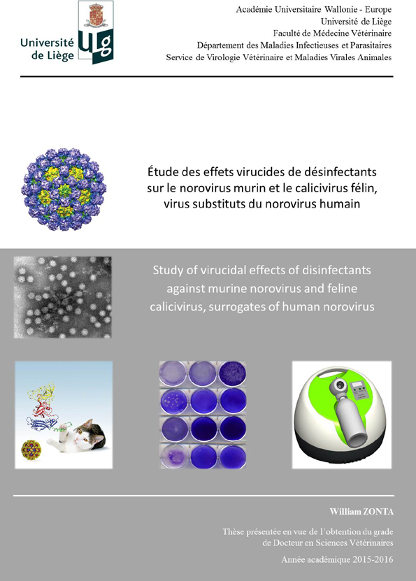
- Title: Study of virucidal effects of disinfectants against murine norovirus and feline calicivirus, surrogates of human norovirus
- Date of the defence: 30th of August 2016
- Academic year: 2015-2016
Human noroviruses (HuNoV) are amongst the leading causes of acute non-bacterial gastroenteritis in humans and annually constitute a global public health problem involving numerous people with great medical, sanitary and economic consequences. The effects of disinfectants against viruses and more specifically against highly resistant viruses such as HuNoV in the environment are little known and data, sometimes empirically obtained, originate from effects observed with bacteria or enveloped viruses.
The aim of this thesis is to evaluate disinfectant effects against HuNoV. Since in vitro culture of HuNoV remains difficult, genetically and morphologically similar viral surrogates, are utilised as indirect tools to obtain data about the virucidal effects of disinfectants. The murine norovirus (MNV) and feline calicivirus (FCV) are two frequently used HuNoV surrogates, for which an in vitro cell culture system and a plaque assay protocol exist.
In the first study, the virucidal effect of seven disinfectants was evaluated in suspension tests and in two carriers tests (stainless steel and the latex gloves), using two parameters, the infectious viral titre and the genomic copy number. This first study demonstrated that three disinfectants were able to induce a viral titre reduction of more than three log10 for the two surrogates in all three kinds of tests. The disinfectants were halogen, an oxidizing agent and a mix composed of quaternary ammonium compounds (QAC), an alcohol and an aldehyde. The three disinfectants also showed a significant effect on the genomic copy number of MNV and FCV in all three kinds of tests. In conclusion, this first study demonstrates that the three disinfectants should be considered as disinfectants with likely efficiency against the HuNoV and that their use should be recommended for disinfection of surfaces or materials potentially contaminated by the HuNoV.
The use of touchless disinfection technologies is gaining increasing importance due to their automatization (limited reliance on human factors), safe use and beneficial economic impact (staff is available for other work). It has been conclusively demonstrated that the environment and contaminated surfaces of health institutes and food processing industries can be a source of infection and have been involved in HuNoV transmission amongst the population in numerous instances.
H2O2 nebulization is a system that aerosolizes a solution, causing it to form liquid droplets in the air. The goal of the second part of this thesis is to evaluate the effect of nebulization on the two main HuNoV surrogates, MNV and FCV, on two different surfaces, i.e. glass and stainless steel, representing the surfaces typically present in field conditions. The H2O2 nebulization was shown to be efficient, mediating a mean viral titre reduction of both surrogates of more than 4 log10, and should henceforth be considered as virucidal. The reductions of genomic copies were relatively low, confirming that this parameter is not an adequate tool for the determination of virucidal efficiency of a system nebulizing H2O2. In order to reduce the risk of contamination and increase the security of consumers, patients and employees, the nebulization of H2O2 should be recommended for use as a disinfection system, complementing conventional disinfection processes in places potentially contaminated by HuNoV.
After the evaluation of the effects of several disinfectants against the MNV and the FCV on the viral titre and the genomic copy number, investigation of the effect of disinfectants was continued by exploring the effect of these disinfectants on the binding step of MNV on RAW 264.7 cells. The aim of this third study was to identify whether the binding step, the first step of the viral replicative cycle, was modified after the use of disinfectants on MNV and whether this mechanism explained the virucidal effect observed in the previous studies. Via epifluorescent microscopy analysis and an indirect immunofluorescence assay, viral attachment was not observed after contact of MNV with alcohol (ethanol and isopropanol), H2O2, chlorhexidine, benzylammonium-chloride (BAC), didecyldimethyl-ammoniumchloride (DAC) at a concentration of 0.08 % or glutaraldehyde. Viral attachment was still partially observed after contact of MNV with the sodium hypochlorite or DAC at a concentration of 0.04 %. All tested disinfectants partially or totally inhibit the binding step, probably due a modification or a destruction of the MNV capsid.
In conclusion, any disinfectants utilized in a disinfection process should include either a halogen product, oxidizing agents or a mix of QAC with an alcohol and an aldehyde in order to guarantee a virucidal effect against viruses such as the HuNoV. The nebulization of hydrogen peroxide should be employed both preventively and in the course of HuNoV outbreaks in places with HuNoV contamination. Amongst different mechanisms of action of virucidal disinfectants, one mechanism against MNV could be the inhibition of cellular receptor binding via structural modification of the viral capsid.
CloseZonta W., Mauroy A., Farnir F., Thiry E. Comparative virucidal efficacy of seven disinfectants against murine norovirus and feline calicivirus, surrogates of human norovirus. Food Environ. Virol., 2016, 8, 1-12, doi:10.1007/s12560-015-9216-21.
Zonta W., Mauroy A., Farnir F., Thiry E. Virucidal efficacy of a hydrogen peroxide nebulization against murine norovirus and feline calicivirus, surrogates of human norovirus. Food Environ Virol., 2016, doi:10.1007/s12560-016-9253-5.
CloseDamien THIRY
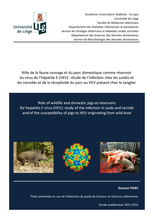
- Title: Role of wildlife and domestic pigs as reservoirs for hepatitis E virus (HEV): study of the infection in suids and cervids and of the susceptibility of pigs to HEV originating from wild boar
- Date of the defence: 18th of December 2015
- Academic year: 2015-2016
The zoonotic transmission of hepatitis E virus (HEV) is of special concern, particularly in high income countries were waterborne infections are less frequent than in developing countries. High HEV seroprevalences can be found in European pig and wild boar populations. The aims of the first study of this thesis were to obtain prevalence data on HEV infection in swine in Belgium and to phylogenetically compare Belgian human HEV sequences with those obtained from swine. Sampling was carried out in the pig serum banks made by the regional animal health laboratories in Belgium between September 2010 and October 2011. In total, 420 serum samples of fattening pigs aged less than 6 months were used for virological studies and 420 serum samples of lactating sows for serological testing. The presence of HEV-specific antibodies was demonstrated by ELISA. An individual seroprevalence of 73 % (95 % CI 68.8-77.5) was found in Belgium. The individual seroprevalence was significantly different between the two regions (Chi2 = 4.83; 1 degree of freedom (df); P = 0.03): 66 % (95 % CI 56.6-74.2) in the Walloon Region and 76 % (95 % CI 71.1-81) in the Flemish Region. Moreover, 93 % (95 % CI 89-100) of the tested herds were found to contain at least one seropositive pig. A collection of 98 pig sera already tested by ELISA was further analysed by a Western blot (WB) against IgG or IgM. These data were obtained in order to use WB as a reference test in a ROC curve analysis. Therefore the ELISA cut-off value was re-evaluated by the ROC curve analysis and different scenarios were analysed. Whatever the scenario, the seroprevalence remained high, from 58 to 77 % for the ELISA seroprevalence and from 69 to 81 % after confirmation by WB. In addition to the investigation of the high HEV seroprevalence in pigs, the risk of zoonotic transmission of the infection was approached by comparing viral sequences identified during this study in pigs and humans in Belgium. Nine human positive serum samples, from the bank of serum of hepatitis E cases of the Belgian National Center for viral hepatitis, obtained between 2009 and 2011, were available in sufficient amount and used for genetic comparison with swine samples. Four out of 420 pig sera were detected positive for HEV RNA. All sequences from the 4 positive pig sera belonged to genotype 3, subtype f. Eight sequenced human HEV fragments belonged to genotype 3 (7 subtypes f and 1 e) and one to genotype 1. These results indicate the possible zoonotic potential of HEV in Belgium. In addition, the existence of other HEV genotypes or subtypes circulating in pigs in Belgium cannot be excluded, as highlighted by the detection of genotype 4 subtype b in a previous survey (Hakze-van de Honing et al., 2009).
In Europe and more accurately in Belgium, the wild boar population is in constant increase with a population size estimated to be more than 25000 heads in 2012 in the Walloon Region (16903 km2). The red and roe deer bag statistics were respectively 5300 and 14 400 in 2012 in the same area. In Belgium, four-fifths of the forests are in the Walloon Region and this zone has a wooded area of 4952 km², representing one third of its total area. Moreover, this region contains also a high density of human population (210 people /km²). The aims of the second study of this work were to obtain prevalence data on HEV infection in wild fauna in Belgium and to phylogenetically compare Belgian human and swine HEV sequences with those obtained from wild boars and cervids. Sampling of sera and livers of wild boars was made during the hunting season from September 2010 to February 2011. A total of 383 sera from wild boars over 6 months of age were selected for serology. For the virological study, all the samples available from young wild boars (less than 6 months of age) were used: 69 sera and 61 livers. Sampling of sera and livers from cervids was also made by the Walloon wildlife surveillance network during the hunting season from October 2012 to December 2012; 189 and 235 sera of respectively red deer and roe deer were collected for serological analysis. For the virological analyses, 84 and 68 sera as well as 29 and 27 livers from respectively red and roe deer were sampled. An overall apparent seroprevalence of 34 % (95 % CI 29.71-39.46) was found in wild boars, of 1 % (95 % CI 0-2.4) in red deer and 3 % (95 % CI 0.8-4.2) in roe deer. In order to assess the ELISA screening prevalence, WB analyses were performed. The data obtained by the WB analysis in wild boars have been used for the conception of a ROC curve analysis with WB as reference test and different scenarios were analysed. Seroprevalence remained high whatever the scenarios in the wild boar population. Indeed, it was from 27 to 64 % for the ELISA screening prevalence and from 34 to 42 % after confirmation by WB. In wild boars, 4 out of 69 sera and 4 out of 61 livers were detected as positive for HEV RNA. All sequences obtained from sera belonged to HEV genotype HEV-3, three to subtype 3f and one to 3c according to the classification of Lu et al. (2006). Comparison with the human and swine strains belonging to genotype HEV-3 showed that most of these sequenced fragments also belonged to subtype 3f. HEV RNA was detected in one out of 29 livers from 109 red deer, it belonged to genotype 3f. No HEV RNA was detected in red and roe deer sera. Using a multivariate logistic regression, a significant effect of age was observed: young animals were less seropositive in comparison with adult animals as reference. A significant effect of density was also observed. Wild boar can be considered as a host reservoir of the virus in Belgium. However, the low prevalence in deer disregards these species as reservoir contrarily to the epidemiological role played by them in other countries. This evidence needs further investigation in order to determine in which situation deer can serve as a reservoir. These results also raise the question of the dynamics of HEV infection between wild fauna, domestic pigs and humans.
An increasing number of animal species have been recognized as susceptible to HEV. Direct zoonotic transmission to human beings, followed by symptomatic infection, has been documented several times as in the case of uncooked meats from wild boar, deer and pigs infected with HEV. The particular epidemiological role of wild boars in the HEV transmission route has recently been investigated. The aim of the third study was first to investigate the early consequences of pig infection with a wild boar HEV strain (WbHEV) inoculated by intravenous route and second to observe the infection pattern of a WbHEV strain, a WbHEV strain previously passed in swine, and a SwHEV strain after oral inoculation. A HEV-3 subtype f isolated from the livers of two young wild boars hunted in the same Belgian forest during the same hunting and phylogenetically similar for the analysed sequences was used for the production of inocula for the intravenous and the oral infection experiments, respectively. In the intravenous infection experiment, the piglets were divided in two groups. Group 1 included three piglets (A, B and C) inoculated with a wild boar HEV strain (WbHEV). Group 2, representing a negative control, was composed of two piglets (D and E) inoculated with the HEV-free liver homogenate. In the oral infection experiment, 12 piglets were divided in four groups. Group 3 included three piglets inoculated with a swine HEV strain (SwHEV); group 4 included three piglets inoculated with a wild boar HEV strain previously passed in swine; group 5 included three piglets inoculated with a wild boar HEV strain (WbHEV); and group 6 was composed of three piglets inoculated with HEV-free liver homogenate representing a negative control. After intravenous inoculation, HEV RNA was detected in serum, bile, liver, spleen, duodenum, jejunum, colon, lung, gastro-hepatic lymph nodes and faeces in all group 1 piglets. Pigs from both groups remained seronegative until the end of the experiment and hepatic enzymes remained within the normal range. Furthermore, no clinical signs were observed. After oral inoculation, HEV RNA was detected in serum, bile, liver, gastro-hepatic lymph nodes and faeces in groups 3, 4 and 5. Most of HEV inoculated pigs became seropositive at day 15 post-inoculation and one Sw-WbHEV inoculated pig became seropositive at day 12. Hepatic enzymes remained within the normal range. Furthermore, no clinical signs were observed. In both oral and intravenous infection experiments, HEV inoculated pigs showed infiltration of lymphocytes, plasmocytes, eosinophils in the portal areas, int erlobular septa and sinusoids. Additionally parenchymal aggregates of lymphocytes, plasmocytes and macrophages could be noticed in parenchyma of the livers. No histopathological differences were observed between the pigs of both groups in duodenum, jejunum and colon samples.
In conclusion, domestic pigs and wild boars can be considered as reservoir hosts for HEV in Belgium. However, contrarily to the apparent epidemiological role of cervids in other countries, the low seroprevalence data obtained in Belgium suggest they are accidental hosts. The results of experimental infections reinforce the putative role of wild boars in the transmission of HEV in pigs. The oral inoculation model better mimics the natural route of infection than the intravenous model and shows a limited spread of the virus in the organism, a low viremia and low levels of the virus in the organs. These points should be considered when assessing the risk of human infection with HEV in the context of the consumption of pork products.
CloseThiry D., Mauroy A., Saegerman C., Thomas I., Wautier M., Miry C., Czaplicki G., Berkvens D., Praet N., van der Poel W., Cariolet R., Brochier B., Thiry E. Estimation of hepatitis E virus (HEV) pig seroprevalence using ELISA and Western blot and comparison between human and pig HEV sequences in Belgium. Vet. Microbiol.., 2014, 172, 407-414.
Thiry D., Mauroy A., Pavio N., Purdy M.A., Rose N., Thiry E., de Oliveira-Filho E. Hepatitis E virus and related viruses in animals. Transbound. Emerg. Dis., 2015, doi:10.1111/tbed.12351.
Thiry D., Mauroy A., Saegerman C., Licoppe A., Fett T., Thomas I., Brochier B., Thiry E., Linden A. Belgian wildlife as potential zoonotic reservoir of hepatitis E virus. Transbound. Emerg. Dis., 2015, doi:10.1111/tbed.12435.
Thiry D., Rose N., Mauroy A., Paboeuf F., Dams L., Roels S., Pavio N., Thiry E., Productive infection in pigs experimentally inoculated intravenously or orally with a wild boar hepatitis E virus (HEV), a swine HEV and a wild boar HEV previously passed in swine. Submitted.
CloseMaria Ana DE LA GRANDIERE DE NORONHA COTTA
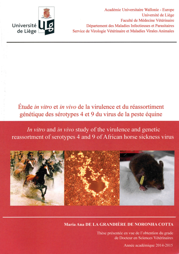
- Title: In vitro and in vivo study of the virulence and genetic reassortment of serotypes 4 and 9 of African horse sickness virus
- Date of the defence: 29th of September 2015
- Academic year: 2014-2015
Vector-borne diseases are a major threat for public and animal health. African horse sickness virus (AHSV) has a segmented genome composed of ten double-stranded RNA segments and belongs to the family Reoviridae and genus Orbivirus. The virus has nine known antigenically distinct serotypes and is transmitted by culicoides biting midges, principally Culicoides imicola. African horse sickness causes severe morbidity and mortality of up to 95 % in horses leading to severe economic losses. The establishment of an experimental mouse model is needed for the investigation of the pathogenesis of this infection, the study of its virulence and the efficacy of vaccines. Three mouse models, interferon-α receptor knock-out mice (IFNAR -/- or A129 KO) and immunocompetent mice (Balb/C and 129 WT), were tested. The virus used for inoculation of mice belongs to the two serotypes, serotype 4 and 9, which have been known to cause epidemics in Europe, The virus was inoculated via subcutaneous route (SC) and/or intranasally (IN). Samples of whole blood were taken for both infected and knock-out mice at regular intervals. Organs (liver, spleen, kidney, lung and brain) were sampled at the end of the experiments, when the most affected mice were euthanized. All these samples were tested by a qRT-PCR, targeting African horse sickness genome segment 7, the most conserved segment between serotypes. Both serotypes (4 and 9) of African horse sickness were detected by qRTPCR until three weeks post-infection (corresponding with the end of the experiments) in blood from SC infected mice. Serotype 4 showed a higher peak of RNAemia than serotype 9. The peak of RNAemia was measured between days 2 and 4 post-infection for the three strains of mice. No RNAemia was detected in blood from IN infected mice during the 3 weeks of the experiment. The setting up of this mouse model has developed a tool for efficient in vivo study to characterize the in vivo virulence of this virus, to monitor the evolution of viral populations during in vivo replication cycles.
In the second study, genetic reassortment for AHSV was investigated. First, in vitro coinfections were performed and reassortant viruses between AHSV serotypes 4 and 9 were generated. Despite a slower viral growth curve for serotype 9 in comparison to serotype 4, dominance of serotype 9 over serotype 4 was observed during coinfection assays and required adaptations of the conditions for coinfections assays. Genetic characterization of the six reassortant viruses by qRT-PCR and RT-PCR showed multiple segment exchanges and a genome constellation per serotype was demonstrated. Three reassortant viruses were selected in order to study their virulence in vivo in comparison with the virulence of the parental strains in Balb/C mice. A higher virulence compared to the parental viruses and other reassortant viruses was observed for one reassortant virus. A correlation between severity of clinical signs and virus detection in blood and organs was found. These data support the modification of virulence by genetic reassortment with possible consequences in virus epidemiology.
In conclusion, in the first part of this thesis, an experimental mouse model was developed. It has proven its effectiveness by its use in the assessment of the virulence of reassortant viruses generated during the second part of the thesis. All the reassortant viruses from this study possess segments 2 (VP2) and 6 (VP5) originating from different parental viruses. This result shows the limitations of conventional serotyping methods which focus exclusively on the identification of segment 2 and therefore do not allow the detection of reassortant viruses.
CloseDe la Grandière A., Dal Pozzo F., Tignon M., Zonta W., Thiry D., Mauroy A., Caij A.B., Saegerman C., Thiry E. Study of the virulence of serotypes 4 and 9 of African horse sickness virus in IFNAR -/-, Balb/C and 129 Sv/Ev mice. Vet. Microbiol., 2014, 174, 322-332.
CloseElisabeth MATHIJS
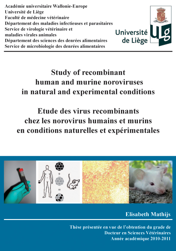
- Title: Study of recombinant human and murine noroviruses in natural and experimental conditions
- Date of the defence: 12th of September 2011
- Academic year: 2011-2012
Noroviruses (NoVs) are among the most important causes of both sporadic cases and outbreaks of gastroenteritis in humans of all ages and are responsible for approximately 90% of epidemic non-bacterial outbreaks of gastroenteritis in industrialised countries. In Belgium, NoVs have been identified as the first cause of foodborne gastroenteritis outbreaks before Salmonella spp and Bacillus cereus. Transmission routes are multiple and outcomes of NoV infections can be particularly severe in health care settings due to the presence of vulnerable patients with underlying severe illnesses. NoVs have been detected in a broad variety of animal species (Scipioni et al., 2008a), raising important, yet partly unanswered, questions about the possibility of zoonotic transmissions and the existence of an animal reservoir for NoVs.
NoVs are highly swift viruses and characterised by great genetic variability that can be attributed to two main evolutionary forces: point mutation and recombination. Both mechanisms allow NoVs to continuously generate mutant genomes. The study on NoV biology including recombination has long been hampered by the lack of cell culture systems and small animal models. Consequently, the discovery of cultivable NoVs that naturally infect laboratory mice gave new perspectives in NoV research. Despite the description of numerous human and animal recombinant NoVs by phylogenetic analysis (Bull et al., 2007), no experimental evidence of NoV recombination after cell culture co-infection is available yet.
The aim of this thesis was to evaluate the importance and the potential implications of NoV recombination on the evolution of NoV in natural conditions and in experimental conditions.
NoVs found associated with foodborne suspected gastroenteritis outbreaks reported to the Belgian Scientific Institute of Public Health between December 2006 and December 2010 were systematically characterised. Therefore, we established a genotyping procedure based upon phylogenetic analyses of the partial sequences of the polymerase and the capsid genes.
A second part was dedicated to the in vitro creation of NoV recombinants after the set up of the murine model. Molecular methods allowing the distinction between two parental wild-type MNVs in different regions of the genomes were implemented in order to detect hybrid genomes among the progeny viruses of co-infections. Furthermore, the consequences of recombination on the biological properties of the recombinant viruses in comparison with the parental ones were assessed in vitro and in vivo.
Among all foodborne suspected outbreaks, NoVs were implicated in 11.8% of the cases and responsible for 34.5% of the persons reported ill. Genogroup 2 (GII) NoVs predominated widely (90.4% of all outbreaks) and GII genotype 4 (GII.4) was detected in 16 of the 28 (57.1%) typed outbreaks. GII.4 2006 variants were repeatedly detected circulating with variants 2007 and 2008 until 2009 before being replaced by a novel variant GII.4 2010 the year after. The latter variant constituted the only GII.4 variant circulating in 2010 and was involved in 8/18 typed outbreaks reported in the winter 2009-2010. Phylogenetic analyses allowed the identification of 5 different combinations of NoVs recombinants associated with 14 outbreaks; four novel GII intergenotype and intersub-genotype recombinants (GII.e/GII.3, GII.g/GII.1, GII.4 2006b/GII.4 2007 and GII.4 2010/GII.4 2010b) were detected along with 1 previously published recombinant (GII.e/GII.4 2007). For all the recombinants, the recombination cross-over was located at the junction of the genes that code for the polymerase and the capsid proteins.
Using the murine NoV (MNV) model, we investigated recombination between two wild-type MNV strains co-infecting mouse monocyte and macrophage cell line (RAW 264.7). A PCR-based genotyping tool capable of discriminating between parental viruses allowed the detection of a viable chimeric virus (Rec MNV) among the progeny viruses. Genetic analysis confirmed the Rec MNV genome to be a chimera created by a homologous recombination event located at the overlap between the ORFs coding for the polymerase and the capsid proteins. In comparison with the parental viruses, Rec MNV showed distinct multiplication kinetics and produced significantly smaller plaques in cell culture. After per oral infections of immunocompetent Balb/c mice with 5.106 plaque forming units of parental viruses and Rec MNV, Rec MNV infected mice showed lower body weight losses at days 2 and 3 post-infection (2-3 dpi) compared to those infected with the parental viruses. Rec MNV was more rapidly cleared from faeces because, contrary to what was observed for the parental viruses, no detectable Rec MNV viruses were found at 3 dpi. Meanwhile, virus titres observed for Rec MNV in faeces and in different organs (small intestine, spleen, mesenteric lymph nodes, left lung) at 2-3 dpi did not differ much from those observed for the parental viruses. Also, the presence of detectable levels of viruses in all organs analysed suggest that, similarly to the parental viruses, Rec MNV can induce a systemic infection and disseminate beyond organs associated with the digestive tract.
The study of NoVs implicated with suspected foodborne gastroenteritis outbreaks in Belgium over a 4-year period highlight the importance of NoVs for public health in our regions. NoVs circulating during this period exhibit great genetic variability due to point mutations and recombination events. The GII.4 2006 until then predominantly present across the globe was successfully displaced by the newly emerging GII.4 2010 variant. Similarly, GII.b recombinants previously considered as endemic on the European continent seem to have been substituted by the nascent recombinants GII.e in 2009 and GII.g in 2010. Among these recombinants, two novel recombinant combinations (GII.e/GII.3 and GII.g/GII.1) have been characterised. Our results also suggested that part of the viruses of the highly successful GII.4 lineage were in fact mosaics of former GII.4 NoVs suggesting recombination might play a role in the divergent evolution of GII.4 NoVs. The emergence of new GII.4 variants together with GII recombinants could explain the explosive increase in reported cases of gastroenteritis that was observed in 2008 and 2010.
The high genetic diversity of NoVs plays a key role in the difficulty to implement efficient prophylactic and therapeutic measures. Consequently, the determination of NoVs predominantly involved in large and cost-effective gastroenteritis outbreaks will allow targeting the development of these procedures on the most prevalent strains. Moreover, taking into account that NoVs undergo point mutations and recombination, the availability of regularly updated vaccines will require the assessment of contemporaneously circulating strains.
Through the use of the MNV model, the present work offers the first experimental evidence of NoV recombination after the co-inoculation in cell culture of two distinguishable MNV strains. The obtaining of a recombinant in permissive conditions without any pressure of selection suggests that the phenomenon is not rare. Nevertheless, different adaptations could be foreseen in order to use the model for the study of NoV recombination. Similarly to what was observed for recombinant HuNoVs in natural conditions, the localisation of the crossing-over at the ORF1-2 overlap with the identification of a stem-loop structure at the anti-genomic strand is in line with the recombination model proposed by Bull and collaborators (2007). This model proposes that the exchange of genetic material occurs during the switch of the polymerase from one genomic strand to the other during RNA negative strand transcription into positive strand RNA.
The evaluation of the biological characteristics of Rec MNV in comparison to the parental viruses both in vitro and in vivo suggests that recombination can result in the creation of chimeric viruses whose properties differ from the parental ones. Its consequences could have a major impact on NoVs evolution like the creation of highly pathogenic viruses or viruses with broadened host ranges. Indeed, the adaptation of a virus in a novel host by point mutations could take years whereas recombination can quickly generate the required genetic combinations.
This thesis allowed the implementation of an in vitro and in vivo NoV study model in our laboratory. The results obtained both in natural conditions for HuNoVs and in experimental conditions for MNVs indicate that recombination, along with point mutation, seem to be highly implicated in the evolution of NoVs. Thus, the understanding of these phenomena will be crucial if therapeutic and prophylactic measures are to be implemented for NoV infections.
CloseMathijs E., Denayer S., Palmeira L., Botteldoorn N., Scipioni A., Vanderplasschen A., Thiry E., Dierick K. Novel norovirus recombinants and of GII.4 sub-lineages associated with outbreaks between 2006 and 2010 in Belgium. Virology Journal, 2011, 18;8: 310.
Mathijs E., Muylkens B., Mauroy A., Ziant D., Delwiche T., Thiry E. Experimental evidence of recombination in murine noroviruses. Journal of General Virology, 2010, 91: 2723-33.
Mathijs E., Dal Pozzo F., Saegerman C., Thiry E. In vivo characterization of an in vitro generated recombinant murine norovirus. In preparation.
CloseShaif ABDULLAH
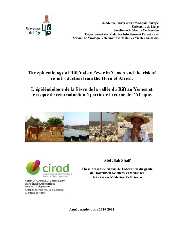
- Title: The epidemiology of Rift Valley Fever in Yemen and the risk of re-introduction from the Horn of Africa
- Date of the defence: 3rd of February 2011
- Academic year: 2010-2011
From 1930 to 2000 Rift Valley Fever (RVF) was limited to the African continent. It is vector borne disease caused by a virus of the genus Phlebovirus, member of the Bunyaviridae family. The main vectors for transmission are Aedes and Culex. In September 2000 it was reported for the first time out of Africa, affecting Yemen and Saudi Arabia. This epidemic opened a new era in the history of RVF. It proved that the virus had the capaciity to affect new zones, different eco-systems and to spread to any area in the world. The outbreak lifted many hypothesis related to its introduction and to the factors associated with the outbreak. Although difficult to evaluate precisely its socio-economic impact was considered to be the heaviest in the modern history of Yemen animal diseases even when compared to the rinderpest outbreak that the country encountered in the seventies because of its zoonotic characteristic. To answer to these hypotheses it was of great importance to study and investigate all the factors associated with the outbreak. Thus after estimating the socio-economic impact of the disease in the world and more specifically in Yemen we studied the descriptive epidemiology of RVF in the first and most affected zone of the outbreak of 2000-2001 (Tihama Wadi Mawr) and then we analysed the socio-economic and environmental factors associated to the outbreak, to finish with the risk assessment of the re-introduction of RVFV from the Horn of Africa through legal animals trade.
The descriptive study showed that at the national level 90% of the RVF cases were in the plain of Tihama coast, Hodiedah, Hajjah and Sadah governorates, the majority of the villages being located around the main canals of Wadi Mawr at an altitude < 300 m.
Environmental as well as socio-economic factors likely to play a role in RVF transmission in Yemen were highlighted with the study of the period 1997-2007 in the country. As in previous RVF outbreaks in neighbouring countries in the Horn of Africa, the year 2000 presented above-normal vegetation index values, which reflected important precipitations, for both rainy seasons (the first occurring between March and May; the second between July and October). These environmental conditions favourable to the vectors populations were found concomitant with a late starting date of Eid-al Kabeer celebration (March) in 2000, related to high hosts (cattle, sheep and goats) densities. According to these criteria, 2000 was considered as an atypical year.
Yemeni Veterinary Services did not declare any RVF outbreak since 2000. Thus, we assumed Yemen free of the disease when assessing the risk of introduction of RVF into Yemen via the legal trade of small ruminants from the Horn of Africa (Kenya, Somalia, Djibouti, and Ethiopia). After precisely describing the routes and volume of trade from the Horn of Africa to Yemen, the pathway and different scenarios for introduction were developed following the OIE risk assessment method. A matrix of likelihood combinations including four possible levels (very low, low, medium, high) was built and used to combine likelihood of events.
The overall probability of introduction was assessed very low to medium depending on the period of the year and most likely to occur via ovine males exported during festival periods that change depending of the year considered. The uncertainty was considered to be low.
The socio-economic impact although difficult to estimate was shown to be dramatic as RVF affects all the chain of life in particular of those associated to livestock and animal products trade.
Despite the dramatic impact of the outbreak of RVF in 2000 but it had the advantage to draw the attention of decision makers, of international organizations and of local veterinary services on the importance of livestock diseases and their possible effects on human health and national economies. Veterinary education also improved significantly in Yemen. It enhanced the epidemiologist’s skills, disease surveillance in general and the cooperation between human health care people and veterinary services. A national P3 laboratory should also soon open with the help of the IAEA and the FAO and Al-Mukkah quarantine could be modernized and extended in the near future.
Regional collaboration and improvement of knowledge on animal trade but also field studies related to the disease and its entomological features are seen as compulsory to hope improving the prevention and control of RVF.But the best way and strategy for prevention of Rift Valley Fever in Yemen as well as in the world is to develop more efficient surveillance and control tools to implement coordinated regional monitoring and control programmes.
CloseAbdo-Salem S., Peyre M., Waret A., Bonnet P., Al-Qasadi M., Saeed K., Thiry E., Roger F., Chevalier V. A review of the socio-economic impact of the Rift Valley fever with a special focus on the Horn of Africa and the Arabic Peninsula. Epidemiology and Infection, accepted for publication.
Abdo-Salem S., Gerbier G., Bonnet P., Al-Qadasi M., Tran A., Al-Eryani G., Roger F. 2006. Descriptive and spatial epidemiology of Rift valley fever outbreak in Yemen 2000-2001. Annals of the New York Academy of Sciences, 1081: 240-242.
Abdo-Salem S., Tran A., Grosbois V., Gerbier G., Al-Qadasi M., Saeed K., Etter E., Thiry E., Roger F., Chevalier V. Can environmental and socio-economic factors explain the recent emergence of Rift Valley Fever in Yemen, 2000-2001? Journal of Vector Borne and Zoonotic Diseases, accepted for publication.
Abdo-Salem S., Waret-Szkuta A., Roger F., Olive M-M, Saeed K., Chevalier V. 2010. Risk assessment of the introduction of Rift Valley Fever from the Horn of Africa to Yemen via legal trade of small ruminants. Tropical Animal Health and Production 2010; published online http://dx.doi.org/10.1007/s11250-010-9719-7.
CloseMaria BARRANDEGUY
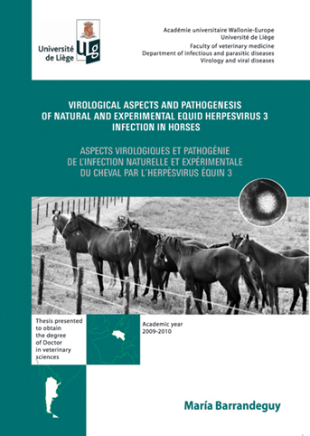
- Title: Virological aspects and pathogenesis of natural and experimental equid herpesvirus 3 infection in horses
- Date of the defence: 14th of September 2010
- Academic year: 2009-2010
Equine coital exanthema (ECE), caused by equid herpesvirus 3 (EHV-3), is a contagious venereal disease characterised by the formation of painful papules, vesicles, pustules and ulcers on the external genitalia of both mares and stallions. EHV-3 is an alphaherpesvirus, distinct from the other equine herpesviruses, endemic in most horse breeding populations worldwide. EHV-3 is primarily transmitted through coitus, although there is also evidence supporting the possibility of non-coital spreading through infected fomites and contacts other than coitus. The infection does not usually result in systemic illness. Epidemiological observations and serological monitoring suggest the existence of latently infected animals from which EHV-3 is periodically reactivated and transmitted to cohorts but latency of EHV-3 has not been formerly demonstrated.
The negative impacts of ECE on equine breeding enterprises are the forced, temporary disruption of the mating activities of mares and stallions, the additional care and supportive treatment in affected horses, and the risk of virus spread by either fresh or frozen semen as well as by artificial insemination and embryo transfer practices. In intensively managed stud operations, which have heavily-scheduled breeding dates for thoroughbred stallions, breeding disruptions may translate into significant end-of-season decreases in the number of entries into the mare book of affected stallions. Also, delayed foaling dates and/or reduced pregnancy rates may occur in mares that miss breeding opportunities due to the disease. Similarly, in the face of an ECE outbreak in artificial insemination and embryo transfer centres, both donor and recipient affected mares show such discomfort that they are reluctant to be inspected, inseminated or transferred, with the consequent loss of opportunity to become pregnant. The additional time and necessary precautions required to manage the donor and receptor mares due to the presence of the disease also have a substantial negative impact.
Because ECE is a not a notifiable disease and the diagnosis is made on the basis of typical clinical signs, most cases and outbreaks of ECE remain unnoticed and its true prevalence and economic impact is difficult to assess and is probably underestimated. Therefore, as several aspects of EHV-3 infection are largely unknown and it has severe economic consequences to the horse industry, the general aim of this doctoral study was to increase the knowledge about the biology of EHV-3 infection. The specific objectives were to investigate the iatrogenic transmission of infection and to set up a protocol for experimental reproduction of the disease, to study the reactivation and re-excretion patterns from latency, to evaluate the epidemiological importance of subclinical infections, and to hypothesise about the ECE economic consequences in the current context of the equine industry.
During the occurrence of an outbreak of ECE in an embryo transfer centre, approximately 32% (n=35) of the donor mares and 25% (n=125) of the recipient mares showed typical ECE lesions around the anus and on the perineal skin, discomfort, and anorectal lymphadenopathy. EHV-3 was detected in 7 (58%) of the affected mares and specific antibodies in 23 (88%) of the convalescent mares. Since no natural breeding had taken place on the affected mares, it could be hypothesised that the virus spread was a consequence of contamination by means of the gloves or the ultrasonography scanner used. Lymphadenopathy provides a new concern associated with ECE.
EHV-3 was isolated from nasal swabs obtained during an outbreak of unilateral rhinitis affecting approximately 40 out of 2000 thoroughbred horses. The fact that an endoscopic examination had been performed in the week previous to the onset of the lesions to evaluate the respiratory tract function was a common finding in all the horses affected. EHV-3 was demonstrated as the etiologic agent of the unilateral rhinitis observed in those 40 thoroughbred horses. The endoscope used for respiratory tract examination was identified as the most likely cause of the spread of the infection. This case is an example of non-genital iatrogenic transmission and reinforces the importance of strict application of hygienic measures in order to reduce the risk of spread of infectious diseases.
The virus isolated from this field outbreak as well as that isolated from others were characterized by means of restriction endonuclease (RE) fragment patterns, plaque size and gG gene partial nucleotide sequencing. In the 25 isolates included in the study, different RE patterns were found: two with BamHI (one of them identical to the one of the reference strain), two with Hind III (both different from the one of the reference strain) and one with Eco RI (different from the one of the reference strain). The plaque size was homogeneous between the isolates, and 1.64 and 2.88 times larger than that of the reference strain. Three base substitutions in the gG gene were found at positions 904, 1103 and 1264, which resulted in strains CAT (Australia), AAT (the United States and Brazil), CAG (Argentina) and ACT (Argentina). The RE pattern and the nucleotide sequence of the gG gene obtained revealed that there are genetically distinguishable EHV-3 strains in circulation. Not only the RE patterns, as previously described, but also the nucleotide sequence of the gG gene, could be useful tools for epidemiological studies. The biological implications of these changes are still unknown.
Two sets of experimental infection with EHV-3 were carried out under controlled conditions. In the first experiment, two seronegative mares were topically inoculated in the vagina and perineal area with EHV-3. The same protocol was followed in the second experiment in two seronegative and two seropositive mares (the mares which had been included in the first experiment were used six months later). Clinical samples consisted of swabs from the vagina and perineal area, and blood samples were obtained for virological, serological and haematological studies. A scoring system was designed and used for daily clinical evaluation and rectal temperature records from each mare. Neither hyperthermia nor haematological changes were recorded in the mares analyzed. Typical ECE lesions were observed in seronegative animals: the clinical score was 172 and 90 (average score: 131) for the mares included in the first experiment and 160 and 92 (average score: 124) for the mares included in the second experiment. Only slight lesions were observed in the seropositive mares, being the clinical score 53 and 41 (average score: 47). Also, differences were detected in the duration and intensity of virus shedding, being 15 and 9 days (duration) and 105 versus 104 (the highest virus load detected) in the seropositive and seronegative mares, respectively.
In one study designed to demonstrate EHV-3 latency and to study reactivation and re-excretion patterns, virus shedding, seroconversion and the presence of a small ECE lesion were observed in one out of two previously naturally infected mares after corticosteroid treatment. EHV-3 was isolated from perineal vaginal swabs of one of the mares, both on day 14 after corticosteroid treatment and along the following 10 days. A small and rounded area of erosion was observed on the left labia of the vulva of the same mare on day 19 after corticosteroid treatment and 5 days after the virus shedding was detected. A significant (four-fold) increase in the antibody titre was found in the mare which shed the virus 28 days after corticosteroid treatment and 14 days after the beginning of virus re-excretion. In concordance with epidemiological observation and serological studies, and in common with other members of the subfamily Alphaherpesvirinae, this study indicates that a state of latency is established after natural infection of EHV-3.
A study was carried out to estimate the prevalence of excretion of EHV-3 under field conditions. The virus was detected in perineal-vaginal swabs by real time PCR and specific antibodies were identified by seroneutralization in 14 (6%) and 105 (48%) respectively of 220 thoroughbred mares without clinical signs at the time of breeding. In order to assess the re-excretion patterns of spontaneous reactivation, two seropositive (presumably latently infected) polo mares were kept in isolation for 11 months. Virological investigations on perineal vaginal swabs obtained on a daily basis revealed re-excretion of EHV-3 on two occasions, 3 months apart (each for a 3-day interval) in one of the mares, and on only 1 day in the other mare. Antibodies against EHV-3 were detected with only slight variation during the entire period in both mares. Clear evidence of the existence of EHV-3 shedders in a healthy mare population under both field and isolation conditions is provided. Furthermore, despite the small number of animals included (only two), the study in mares kept in isolation demonstrated that at least two periods of EHV-3 spontaneous reactivation and re-excretion in the presence of serum antibodies were possible in the same animal in an 11-month interval.
In conclusion, EHV-3 infections and ECE are still a threat for the equine industry. In the present study, EHV-3 was found in several field outbreaks of ECE in thoroughbred breeding farms; the disease was reported by the veterinarians as a true sanitary problem and thus demands additional preventive measures. In addition, EHV-3 was detected as a not rare event in clinically healthy mares, which constitutes the most relevant finding from an epidemiological perspective. ECE is also a sanitary problem of concern for embryo transfer and routine veterinary practices.
The population of EHV-3 latently infected mares which reach up to 50% at the time of breeding deserves special attention. Reactivation of the latent virus is not preventable and those mares can spontaneously reactivate the virus and become a source of infection for highly valuable horses like « shuttle stallions », with the consequent economically negative impact on the equine enterprises.
Finally, there is an important need for finding out additional preventive measures including « pen side » diagnostic tools that allow the detection of subclinically EHV-3 shedding mares in order to segregate them from natural breeding and give them an appropriate antiviral treatment before being covered by stallions.
CloseBarrandeguy M., Perkins J., Mac Donough J., Vissani A., Olguin C., Thiry E. Occurrence of Equine Coital Exanthema in mares from an embryo transfer center. Journal of Equine Veterinary Science, 2010, 30, 145-149.
Barrandeguy M., Ulloa N., Bok K., Fernandez F. Outbreak of rhinitis caused by equid herpesvirus type 3. Veterinary Record, 2010, 6, 178-179.
Barrandeguy M., Vissani A., Miño S., Olguin C., Becerra L., Tordoya S., Thiry E. Characterisation of Equid herpesvirus 3 Argentinean field isolates. Manuscript in preparation.
Barrandeguy M., Vissani A., Olguin C., Barbara G., Valenzuela H., Becerra L., Tordoya M., Miño S., Thiry E. Experimental infection with Equid herpesvirus 3 in seronegative and seropositive mares. Manuscript in preparation.
Barrandeguy M., Vissani A., Olguin C., Becerra L., Miño S, Pereda A., Oriol J.,Thiry E.Experimental reactivation of Equid herpesvirus 3 following corticosteroid treatment. Equine Veterinary Journal, 2008, 40, 593-595.
Barrandeguy M., Vissani A., Pont Lezica F., Salamone J., Heguy A., Becerra L., Olguin Perglione C., Thiry E. Subclinical infection and periodic shedding of equid herpesvirus 3. Theriogenology 2010, doi:10.1016/j.theriogenology.2010.03.14.
Barrandeguy M., Thiry E. Equine coital exanthema-Equid herpesvirus 3: a disease of concern for the equine industry. Veterinary Journal, 2010. (in press).
CloseAxel MAUROY
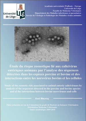
- Title: Study of the zoonotic risk associated to animal enteric caliciviruses by analysis of sequences detected in the porcine and bovine species, and of the interactions between bovine noroviruses and cells
- Date of the defence: 21st of June 2010
- Academic year: 2009-2010
Enteric caliciviruses were detected in humans and animals incoming into food chain such as porcine and bovine species. Caliciviruses have a single stranded, positive, polyadenylated RNA genome. They share properties of high environmental stability, high excretion load and high genetic variation linked to point mutations and recombination. These properties have allowed the formulation of hypothesis about zoonotic transmission or animal reservoir for human strains. The aim of the thesis, composed of four studies, was to investigate the zoonotic risk associated to animal enteric caliciviruses.
In a first study, circulation of both sapoviruses and noroviruses was evidenced by molecular detection in Belgian pig farms. They were detected in both asymptomatic animals or in piglets showing clinical signs of enteritis.
In a second study, bovine noroviruses were molecularly detected in Belgian cattle. Strains phylogenetically related to those of the genotype 2 were predominant. In the same study, seroprevalence against bovine norovirus infection in cattle was investigated by indirect ELISA. Antigens included in the ELISA were virus-like particules obtained in the baculovirus system by expressing the capsid protein of a strain isolated by the laboratory during a previous study. Apparent seroprevalence was high (93.2%), confirming previous results about apparent molecular prevalence in diarrheic calves (7.5%).
In a third study about molecular detection of bovine noroviruses, diagnostic strategy was revised in order to improve the detection of genotype 1 strains and to deal with opportunity of recombination events. Bovine norovirus recombinant strains and also, surprisingly, some sequences genetically related to bovine kobuviruses were detected.
In a fourth study, attachment factors and internalization pathways for genotype 2 bovine noroviruses were studied with an original quantitative method based on flow cytometry analysis. Along with a galactosyl residue that seems to be essential, a sialic acid residue was also showed to be implied in the binding of genotype 2 bovine noroviruses or in a posterior step. Internalisation pathways related to lipid rafts and to macropinocytosis were found.
Together the results have contributed to the analysis of the zoonotic risk associated to enteric animal caliciviruses in the Belgian epidemiological situation. According to these results, the zoonotic risk seems to be low as no sequences genetically related to the human ones were detected. However, some results suggest to maintain a certain degree of vigilance. Indeed, molecular detection showed the co circulation of both bovine and porcine noroviruses in Belgian farms, implicating that hazard exposition exists and could be high in the Belgian epidemiological situation. Morevover, circulation of recombinant strains in the overall population of the bovine norovirus strains implies that this phenomenon was included in the risk assessment. Infection pressures and high prevalences for human and animal strains in a closed epidemiological context as the Belgian one could increase the risk of interspecific recombination. Finally, the sialic acid residue, possibly involved in the binding of genotype 2 bovine noroviruses, and a poorly specific internalisation pathway as macropinocytosis could also favor interspecies barrier crossing.
In the current knowledge, zoonotic risk associated to animal enteric caliciviruses can be communicated as low but a degree of vigilance has to be retained, associated for example to observation of genetic evolution in the populations of human and animal strains.
CloseMauroy, A., Scipioni, A., Mathijs, E., Miry, C., Ziant, D., Thys, C., Thiry, E. Noroviruses and sapoviruses in pigs in Belgium. Archives of Virology, 2008a, 153, 1927-1931.
Mauroy, A., Scipioni, A., Mathijs, E., Saegerman, C., Mast, J., Bridger, J. C., Ziant, D., Thys, C., Thiry, E. Epidemiological study of bovine norovirus infection by RT-PCR and a VLP-based antibody ELISA. Veterinary Microbiology, 2009a, 137, 243-251.
Mauroy, A., Scipioni, A., Mathijs, E., Thys, C., Thiry, E. Molecular detection of kobuviruses and recombinant noroviruses in cattle in continental Europe. Archives of Virology, 2009b, 154, 1841-1845.
Mauroy, A., Gillet, L., Mathijs, E., Thiry, E. Alternative attachment factors and internalisation pathways for GIII.2 bovine noroviruses. Submitted for publication.
Mauroy, A., Scipioni, A., Mathijs, E., Thiry, E. Les norovirus bovins, virus entériques méconnus dans l'espèce bovine. Annales de Médecine Vétérinaire, 2008b, 151, 257-268.
Scipioni, A., Mauroy, A., Vinje, J., Thiry, E. Animal noroviruses. Veterinary Journal, 2008, 178, 32-45.
CloseGuillaume FORTIER
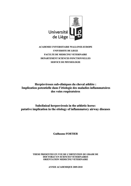
- Title: Study of subclinical herpesvirus infections in the athletic horse in the aetiology of airway inflammatory diseases
- Date of the defence: 12th of December 2009
- Academic year: 2009-2010
Equine gamma-herpesvirus 2 and 5 have been identified worldwide since many years. Their real pathogenic role has still to be determined. On the other hand, many important studies have been performed in order to better understand subclinical diseases of the athletic horse. Several publications pointed out the potential incidence of viruses in lower airway inflammation.
Within the first step of this work, over 700 respiratory fluids were tested in order to detect and to evaluate the presence of equid herpesviruses in these biological samples. Using molecular tools, Equid herpesvirus 2 was found to significantly be the most important among the four herpesviruses detected in tracheal washes of a group of poorly-performing horses. An experimental infection with equid herpesvirus 2 was then conducted on 6 horses and succeeded in reproducing clinical and cytological signs of disease and virus shedding in most biological samples including tracheal washes. The virus shedding was concomitant with ciliocytophthoria of the exfoliated epithelial cells and liquid neutrophilia. Equid herpesvirus 2 can induce a tracheal inflammation and ciliocytophthoria.
This virus could play a role in the new syndrome of tracheal inflammation, and could be an important feature for equine industry.
CloseFortier G., Van Erck E., Pronost S., Lekeux P., Thiry E. Equine gamma-herpesvirus : pathogenesis, epidemiology, and diagnosis. Vet. J., in press, doi: 10.1016/j.tvjl.2009.08.017.
Fortier G., Pronost S., Miszcak F., Fortier C., Léon A., Richard E., Van Erck E., Thiry E, Lekeux P. Identification of equid herpesvirus-5 in respiratory liquids: A retrospective study of 785 samples taken in 2006-2007. Vet. J., 2009, 182, 346-348.
Fortier G., Van Erck E., Fortier C., Richard E., Poitier D., Pronost S., Miszczak F., Thiry E, Lekeux P. Herpesvirus in respiratory liquids of horses: putative implication in airway inflammation and association with cytological features. Vet. Microbiol., 2009, 139, 34-41.
CloseAlexandra SCIPIONI
- Title: Genotypical study of contemporaneous human and bovine noroviruses and development of fast methods for detection and quantification
- Date of the defence: 25th of May 2009
- Academic year: 2008-2009
Noroviruses (NoV), belonging to the family Caliciviridae, are a major cause of epidemic and sporadic cases of highly contagious gastroenteritis in human. Their transmission follows the fecal-oral way and they are a very important cause of human foodborne outbreaks, in particular due to the consumption of bivalve molluscs. Their genome is made of a positive single stranded RNA and they are classified by genetic analysis into 5 genogroups, each of those containing several genotypes.
NoV can not be multiply in a cell culture system. The RT-PCR became the method of choice for their detection in stool samples, food and environmental samples. It is important to have sensitive techniques that can also detect a broad range of NoV. The quantification of the viral load is possible using real-time RT-PCR and is important, not only to estimate the contamination level of a sample, but also to study and characterize the pathogenesis of a NoV infection.
NoV were detected in several animal species, among which the bovine species. These discoveries raised important questions about a zoonotic transmission and the existence of an animal reservoir. The molecular characterization of the two prototypes of bovine NoV, namely Newbury2 virus and Jena virus, revealed that they are genetically close and associated with human NoV. Among the possible hypothesis, animals could be either passive carriers of NoV, or infected in an active way by these viruses, consequently responsible of a zoonosis. The characterization of NoV circulating in human and animal species is interesting in the aim of studying their transmission routes and the possible inter-species transmission.
An important mechanism for NoV evolution is recombination, of great interest in the study of NoV, leading to modifications of the viral genome by exchange of genomic segments. This creates genetic variation and the emergence of new viruses. Indeed, it is well documented that the recombination often occurs among NoV and contributes to the genetic diversity of these viruses as well as to the appearance of new epidemic strains. The prevalence of recombinant strains of NoV could be underestimated by the fact that the characterization of NoV is usually based only on the partial sequencing of the ARN-dependent ARN-polymerase gene only. Whereas ideally various parts of the genome, mainly the ARN polymerase-ARN dependent and the capsid protein, would be sequenced to identify such viruses. It is necessary to determine precisely the role of recombination in the evolution of NoV in order to understand the evolution of the strains and advantages given to some of these strains. The study of this mechanism will allow to better understand the selective advantage observed with some NoV and will help to elucidate the transmission routes of NoV. The study of these two points (zoonotic transmission and recombinant viruses) is very important. Indeed, the cross of species barrier would affect at the same time the epidemiology and the evolution of these viruses, and would also complicate the capacity to develop a vaccine or a treatment. In other animal species, like horses or birds, no NoV was detected to date but these last years, NoV were described in several animal species (dog, lion, sheep). That lets predict a wider range of target species.
This work enters within the scope of the study of transmission routes of NoV with the objectives, after the development of fast and sensitive methods for detection and quantification, to give an explanation to important matters relating to the evolution of NoV and their host range. To begin, the conventional RT-PCR was used in order to detect NoV in human and bovine. Then, a real-time RT-PCR using SYBR Green technology was developed and uses an internal control made up of RNA added in the same tube. This test is able to detect human and bovine NoV belonging to the genogroups I, II and III and makes it possible to make the distinction between NoV and the internal control by the analysis of melting curves. A 10-fold dilution of the samples appeared the method of choice to get rid of inhibition.
In order to confirm the result directly and to allow the quantification of NoV, a real-time RT-PCR using TaqMan technology was developed. It uses an internal control of RNA and also a RNA standard. In a very interesting way, this method can detect human NoV belonging to the genogroups I, II and bovine NoV of the genogroup III. Inhibitions were effectively raised by a 10-fold dilution of the sample or the addition of bovine serum albumin in the RT-PCR mix.
These two real-time RT-PCR showed a higher sensitivity compared to the conventional RT-PCR.
With the objective of understanding the transmission routes of NoV, the situation in Belgium was studied and human and bovine NoV were detected and analyzed using partial sequencing. NoV belonging to different genogroups were detected: GI and GII in human and GIII in bovine. By analysis of the genetic proximity, bovine NoV are close to genotype GIII.2 and human NoV close to various genotypes, but mainly GII.4.
These analyses allowed the identification of two natural recombinants and a co-infection, the latter showed two different genotypes according to the area of the genome being analyzed (polymerase or capsid). The identification of a hot-spot for recombination at the junction of the ORF (open reading frame) 1 and ORF2 confirms the importance of this part of the genome for this phenomenon.
In order to study the evolution of bovine NoV, a strain detected in Belgium was sequence completely (Bo/B309/2003/BE) and compared with the original strain Newbury2 (detected in the 80s).
On one hand, this study made it possible to set up and validate tools allowing the detection and the study of human and animal NoV regarding their pathogenesis, evolution and transmission routes.
On the other hand, based on the database of samples collected during this study, the phylogenetic analysis of NoV detected and characterized goes in the direction of the studies carried out in other countries tending to show that bovine NoV make a group of distinct viruses, different from human NoV. That suggests that bovine NoV do not represent a risk for the human health. Nevertheless, the hypothesis of a zoonotic infection or an animal reservoir cannot be excluded considering the sequence proximity between human and animal NoV and also the close relationship between the human populations and the animal breedings.
The detection of a co-infection and two natural recombinants shows the evolution opportunities of NoV and the importance of a complete analysis of their genome for their characterization.
This work was the first in the field of NoV in the laboratory and has opened the way for other research subjects on NoV and new PhD thesis, especially on the study of NoV-host interaction (NoV in bovine species) and the study of recombination in vitro (murine NoV).
CloseScipioni, A., Mauroy, A., Vinje, J., Thiry, E. Animal noroviruses. Vet. J., 2008, 178, 32-45.
Scipioni, A., Bourgot, I., Mauroy, A., Ziant, D., Saegerman, C., Daube, G., Thiry, E. Detection and quantification of human and bovine noroviruses by a TaqMan RT-PCR assay with a control for inhibition. Mol. Cell. Probes, 2008, 22, 215-22.
Scipioni, A., Mauroy, A., Ziant, D., Saegerman, C., Thiry, E. A SYBR Green RT-PCR assay in single tube to detect human and bovine noroviruses and control for inhibition. Virol. J., 2008, 5, 94.
Scipioni, A., Mauroy, A., Mathijs, E., Ziant, D., Daube, G., Thiry, E. Detection of contemporaneous human and bovine noroviruses in Belgium. Submitted for publication.
CloseJulien THIRY
- Title: Viral diversity and heterologous protection in the cluster of ruminant alphaherpesviruses related to bovine herpesvirus 1
- Date of the defence: 30th of November 2007
- Academic year: 2007-2008
Introduction
Herpesviruses have mainly co-evolved with their hosts for millions of years. Consequently, different related host species may have been infected by various genetically related herpesviruses. Illustrating this concept, several ruminant alphaherpesviruses have been shown to form a cluster of viruses closely related to bovine herpesvirus 1 (BoHV-1). This latter virus, prototype of this cluster, is a major pathogen in cattle associated with various clinical manifestations including infectious bovine rhinotracheitis (IBR) and infectious pustular vulvovaginitis (IPV). IBR is a disease of major economic concern in many parts of the world and especially in Europe, both in European countries where this viral infection has been eradicated and in those where the control of IBR is currently or will be undertaken. The massive use of vaccination allowed the reduction of the number of clinical IBR cases.
However, the existence of alphaherpesviruses closely related to BoHV-1 has to be taken into account in IBR control. The following viruses can be distinguished: bovine herpesvirus 5 (BoHV-5) responsible for severe meningo-encephalitis in calves, bubaline herpesvirus 1 (BuHV-1) inducing a subclinical genital infection in water buffalo (Bubalus bubalis), caprine herpesvirus 1 (CpHV-1) causing systemic disease in young kids and abortion in adult goats, cervid herpesvirus 1 (CvHV-1) responsible of severe ocular syndrome in red deer (Cervus elaphus), cervid herpesvirus 2 (CvHV-2) and elk herpesvirus 1 (ElkHV-1), which induce a subclinical genital infection respectively in reindeer (Rangifer tarandus) and elk (Cervus canadensis). Ruminant alphaherpesviruses related to BoHV-1 are antigenically and genetically closely related to each other. Their common biological properties allow heterologous infection and therefore the crossing of the species barrier. Thus, experimental cross-infection studies show that BoHV-1 is able to infect buffalo, goats, red deer, reindeer and sheep. Inversely, bovines are susceptible to BoHV-5, CpHV-1, CvHV-1 and CvHV-2.
Consequently, the main objective of the present work is dedicated to afford a better knowledge of the interaction between alphaherpesviruses and their ruminant hosts. To meet the objective, two approaches have been developed: the study of the viral diversity aiming to extend both epidemiological and virological data about ruminant alphaherpesviruses related to BoHV-1 and the study of the heterologous protection aiming to protect a minor ruminant species against infections with BoHV-1 related alphaherpesviruses.
Results
Illustrating the issues associated to the distribution of ruminant alphaherpesviruses related to BoHV-1, an original situation has been described in Belgium. In 2001 and 2002, 28.9% of red deer have been detected seropositive to BoHV-1. In absence of contact between cattle and red deer, it has been suggested that a virus related to BoHV-1 was spreading in the Belgian red deer population. Nasal and genital swabs have been collected in hunted red deer and have been inoculated onto bovine kidney cells in order to isolate the viral agent responsible of the seropositivity of Belgian red deer. A viral isolate was detected from nasal sample of a male fawn hunted in the Anlier forest. The analysis of a 443 bp sequence of glycoprotein B demonstrated that the viral isolate was related to ruminant alphaherpesviruses and especially CvHV-1. This red deer alphaherpesvirus was then characterised by comparison with the other ruminant alphaherpesviruses. The viral isolate was antigenically distinct from the other related viruses although it presumably possesses some common epitopes with CvHV-1 and CvHV-2 because a weak reaction was detected in immunofluorescence. The analysis of BamHI and BstEII restriction profiles showed ruminant alphaherpesviruses and especially the viral isolate can be distinguished by their genomic profile. A phylogenetic analysis of these related viruses has been also undertaken. Partial sequence data from UL27 encoding the glycoprotein B (gB) and US8 encoding the glycoprotein E (gE) have been sequenced. This analysis has confirmed that the viral isolate and related viruses constituted a consistent cluster within the subfamily of alphaherpesviruses.
BoHV-5 and BuHV-1 clustered together and are the most closely related to BoHV-1 1.1, BoHV-1 1.2. CpHV-1 is the most diverging ruminant alphaherpesvirus. CvHV-1 is more related to BoHV-1 than ElkHV-1 and CvHV-2 which is the most closely related to CpHV-1. The Anlier isolate clustered with CvHV-1 and the most closely related virus is ElkHV-1. The viral isolate is indeed a different CvHV-1 strain than the CvHV-1 Banffshire 82 strain. Taken together, these data report the first isolation of a ruminant alphaherpesvirus in wild fauna, which has been named CvHV-1 Anlier.
This isolation demonstrates that a ruminant can be identified as positive to BoHV-1 while it is infected with another alphaherpesvirus. In France, cattle purchased from herds so-called “IBR-free status” are sporadically identified as seropositive to BoHV-1. Among other hypotheses, the likely explanation for this “false” seropositivity would be a cross-infection with a related alphaherpesvirus. The most “plausible” virus leading to such diagnosis misinterpretation is CpHV-1 infecting goat. In this context, an epidemiologic study has been performed in order to determine if a CpHV-1 infection was present in France. The analysis of 2,548 serums in BoHV-1 gB blocking ELISA revealed that a ruminant alphaherpesvirus infection was spreading in Corse-du-Sud while no goat was detected positive in Dordogne and Vendée. Taking into account the results obtained in Dordogne and Vendée, a specificity of 100% has been measured for the BoHV-1 gB blocking ELISA. By testing serums from goats experimentally infected with CpHV-1, the sensibility has been calculated to 93.5%. The analysis in BoHV-1 gE blocking ELISA showed that 22.6% of gB-seropositive serums were also gE-seropositive. The BoHV-1 gE blocking ELISA was therefore not able to differentiate between BoHV-1 and CpHV-1 infections. However, the analysis of serums in cross seroneutralisation has strongly identified antibodies against CpHV-1 in gB-positive serums. The presence of CpHV-1 in Corse-du-Sud associated to high prevalence (61.9%) in all analysed flocks extends the number of countries infected with CpHV-1. Moreover, the difference observed between Corse-du-Sud and Dordogne and Vendée suggests that CpHV-1 is more prevalent in Mediterranean regions or countries than in Central or northern Europe.
CpHV-1 therefore appears as the BoHV-1 related virus which has the highest economic concern in Europe. Goat is a species defined as minor and the veterinary medicine industry has no commercial interest toward vaccine development protecting this species against CpHV-1. As BoHV-1 and CpHV-1 are antigenically and genetically related, a live attenuated BoHV-1 vaccine carrying a deletion in the gene encoding gE could protect goats against CpHV-1 infection. The vaccine safety has been assessed by intranasal inoculation of either BoHV-1 virulent strain or gE deleted BoHV-1 vaccine in two groups of two goats. The length of viral excretion and the peak viral titre have been decreased in the immunised group. In order to assess the vaccine efficacy, a group of goats was immunised twice two weeks a part with the vaccine while a control group was kept uninoculated. Four weeks later, immunised and control goats have been intranasally challenged with CpHV-1. A decrease of the peak viral titre was observed in immunised goats. However, the viral excretion lengthened two days more than in control group. It is concluded that this live attenuated gE-negative BoHV-1 vaccine induces a partial cross-protection against a CpHV-1 nasal infection in goats.
In regards to the results obtained in this preliminary experiment, the vaccine has been assessed against a CpHV-1 genital infection which is the natural route of infection in goats. To reach this goal, a group of goats was immunised twice three weeks a part with the gE-negative BoHV-1 vaccine followed by a subsequent CpHV-1 intravaginal challenge. To analyse the safety and the efficacy of this marker vaccine, two groups of goats served as controls: one was immunised with a virulent CpHV-1 and one was kept uninoculated until the challenge. The vaccine did not induce any undesirable local or systemic reaction and goats did not excrete gE-negative BoHV-1. After challenge, a significant reduction in disease severity was observed in immunised goats. Moreover, goats immunised with either gE-negative BoHV-1 or CpHV-1 exhibited a significant reduction in the length and the peak of viral excretion. Antibodies neutralising both BoHV-1 and CpHV-1 were raised in immunised goats. These data show that the intranasal application of a live attenuated gE-negative BoHV-1 vaccine is able to afford a clinical protection and a reduction of virus excretion in goats challenged by a CpHV-1 genital infection.
Conclusions
This study has been performed in order to afford a better knowledge of the interaction between alphaherpesviruses and their ruminant hosts. The antigenic, genetic and genomic relationships existing between BoHV-1 and ruminant alphaherpesviruses have been analysed. It has been demonstrated that these viruses constitute a strong cluster within the subfamily of alphaherpesviruses. The exact knowledge of the evolutionary relationships among these viruses request complete genome sequencing but this analysis is complicated by the large size of herpesvirus genome. Another possibility would be to compare different regions distributed along the genome of each virus as demonstrated by the study of the BoHV-4 history evolution (Dewals et al., 2006). Such analysis could bring interesting data about recombination and transmission events that occurred in the past between herpesviruses and their ruminant species. Nevertheless, the present work gives a contribution to the knowledge of the natural history of the cluster of ruminant alphaherpesviruses related to BoHV-1. Thus, the first isolation of CvHV-1 in a free-ranging red deer linked to the knowledge of the evolution of red deer populations in Europe allowed an interesting hypothesis about the origin of this virus. In regards of the observed prevalence and the presence of red deer over large area, this virus could be responsible of an endemic infection spreading in Europe. This virus may have been introduced after European red deer importation to constitute the initial stocks of Scottish farms. Consequently, CvHV-1 Anlier strain could be the ancestor of the Scottish CvHV-1 Banffshire 82 strain. However, molecular epidemiologic study of several isolates of the two strains is required to firmly assess this hypothesis.
This isolation has demonstrated that misinterpretation of the serologic status of ruminant can be observed; the Belgian red deer was not infected with BoHV-1 but with its own herpesvirus. These difficulties of interpretation are encountered in France in bovines free of IBR and detected later as positive in serology. As BoHV-5 and ElkHV-1 have not been identified in Europe, that there is a very few number of buffalo farms in France, and that CvHV-1 and CvHV-2 transmission to bovine would be exceptional, the only virus that could be responsible of misdiagnosis in France is CpHV-1. Indeed, goats and cattle can be in close contact in some field situations. The results obtained in the second study did not support this hypothesis.
Indeed, CpHV-1 infection is either absent or at a very low prevalence in continental France. Contrarily, a very high seroprevalence of CpHV-1 was measured in the Corse-du-Sud department. In Corsica, the contact between cattle and goats is very limited because flocks and herds are not mixed. Therefore, the transmission of CpHV-1 from goats to cattle would be only exceptional, such cross-infection being very rare, and a BoHV-1 infection in goats is not likely to occur. As the current sampling is restricted to Dordogne and Vendée, a particular attention to CpHV-1 has to be paid in continental France and especially in the Mediterranean region. Such monitoring can be performed with the BoHV-1 gB blocking ELISA which has been validated in the current study.
The CpHV-1 infection is therefore extended to the entire Mediterranean region and appears to be the most relevant ruminant alphaherpesvirus infection besides BoHV-1. In this context, it is necessary to protect goats to reduce the CpHV-1 circulation in the caprine population and economic concerns caused by this infection. The preliminary assessment of the live attenuated BoHV-1 vaccine carrying a deletion in the gene encoding gE has shown that goats could respond to such vaccination. As goat is completely susceptible to BoHV-1, this species could also be used as an experimental model to study the in vivo BoHV-1 properties. A partial efficacy against CpHV-1 nasal infection has been observed. Moreover, the gE-negative BoHV-1 vaccine has been assessed against CpHV-1 genital infection which is the natural route of infection in goats. The intranasal use of a gE-negative BoHV-1 vaccine enabled a clinical protection and a significant reduction of the viral excretion in goats infected genitally with CpHV-1. These results demonstrate that nasal mucosa can serve as an efficient site for the induction of a specific protective response in the genital tract. In humans, several mechanisms are proposed to explain such immune induction involving antibody secreting cells and leading to specific IgA and IgG responses.
Such mechanisms could be involved in the observed response in vaccinated goats but were not analysed in the current study. The biosafety of such cross protection induced with BoHV-1 vaccine against CpHV-1 has also to be considered. Indeed, viruses can be established in a latent state. In bovine, experiments have shown that gE-negative viruses are less reactivated and reexcreted than wild viruses. Consequently, the risk of reactivation and reexcretion in goats is therefore very low. Otherwise, the emergence of recombinant viruses between CpHV-1 and BoHV-1 after cross infection in goats has to be discussed. Among the cluster of ruminant alphaherpesviruses related to BoHV-1, only two recombinant viruses between BoHV-1 and BoHV-5 were isolated, and no recombinant between BoHV-1 and less closely related CpHV-1 was detected in vitro.
Consequently, the cross vaccination described here is likely to be completely safe. Moreover, the use of already licensed vaccine is interesting knowing that the development of new vaccines to protect minor species is of a weak interest for the pharmaceutical industry.
CloseThiry J., Keuser V., Muylkens B., Meurens F., Gogev S., Vanderplasschen A., Thiry E. Ruminant alphaherpesviruses related to bovine herpesvirus 1. Vet. Res., 2006, 37, 169-190.
Thiry J., Tempesta M., Camero M., Tarsitano E., Bellacicco A.L., Thiry E., Buonavoglia C. A live attenuated glycoprotein E negative bovine herpesvirus 1 vaccine induces a partial cross-protection against caprine herpesvirus 1 infection in goats. Vet. Microbiol., 2006, 113, 303-308.
Thiry J., Widén F., Grégoire F., Linden A., Belák S., Thiry E. Isolation and characterisation of a ruminant alphaherpesvirus closely related to bovine herpesvirus 1 in a free-ranging red deer. BMC Vet. Res., 2007, 3, 26.
Thiry J., Saegerman C., Chartier C., Mercier P., Keuser V., Thiry E. Serological evidence of caprine herpesvirus 1 infection in Mediterranean France. Vet. Microbiol., 2008, 128, 261-268.
Thiry J., Tempesta M., Camero M., Tarsitano E., Muylkens B., Meurens F., Thiry E., Buonavoglia C. Clinical protection against caprine herpesvirus 1 genital infection by intranasal administration of a live attenuated glycoprotein E negative bovine herpesvirus 1 vaccine. BMC Vet. Res., 2007, 3, 33.
CloseBenoît MUYLKENS
- Title: Study of the recombination of bovine herpesvirus 1: virulence of recombinant viruses deleted in the glycoprotein E gene and genomic tracing of recombination events
- Date of the defence: 22nd of May 2006
- Academic year: 2005-2006
Bovine herpesvirus 1 (BoHV-1), classified as an alphaherpesvirus, is a major pathogen of cattle. Primary infection is accompanied by various clinical manifestations such as infectious bovine rhinotracheitis (IBR), infectious pustular vulvovaginitis, abortion and systemic infection. When animal survives, a latent infection is established in sensory ganglia. A reactivation stimulus can lead to viral re-excretion. In regards to the significant losses incurred by disease and trading restrictions, several European countries have initiated BoHV-1 control programs based on the use of marker vaccines deleted in the gE gene. These marker vaccines, either inactivated or live attenuated, used together with a serological detection of gE-specific antibody (Ab), allow differentiation between infected and vaccinated animals.
The pathogenicity of BoHV-1 considers the existence of coinfection situation in cattle. Previous data supported the frequent rise of BoHV-1 recombinants in cattle after concomitant nasal infections with two BoHV-1 mutants (Schynts et al., 2003b). The situation is even more complex because of the common use of live attenuated BoHV-1 vaccines injected at the intranasal mucosa. Because the nasal mucosa is the natural portal of entry of wild-type BoHV-1, recombination between a field strain and a gE negative vaccine has been envisaged. Therefore, there is a concern about the virulence of gE negative BoHV-1 recombinants rising from coinfection between a virulent BoHV-1 and a gE negative BoHV-1 vaccine.
Intramolecular recombination is a mechanism of the genetic material exchange closely related to the alphaherpesvirus replication cycle (Thiry et al., 2005). It occurs frequently between strains of herpes simplex virus (Brown et al., 1992; Umene, 1985), varicella zoster irus (Dohner et al., 1988), feline herpesvirus 1 (Fujita et al., 1998), PrV (Glazenburg et al., 1994) and BoHV-1 (Meurens et al., 2004a; 2004b; Schynts et al., 2003b). Several studies have shown that mixed infection with two avirulent strains of the same alphaherpesvirus species can result in a synergistic increase in the severity of disease through the generation of recombinant viruses (Brandt et al., 2003; Javier et al., 1986). Presently, no data are available for the assessment of the risk encountered by gE negative recombinants. The gE negative phenotype, acquired from the vaccine strain, is supposed to guaranty the virus attenuation. However, virulence factors inherited from the wild-type BoHV-1 could enhance the virulence of such recombinants.
Three approaches were developed in this thesis. They aimed at (i) generating gE negative BoHV-1 recombinants, (ii) assessing the virulence of such recombinants, (iii) localizing the recombination that lead to their rise.
The first study was devoted to the generation and the biological characterization of gE negative BoHV-1 recombinants. We decided to produce the gE negative recombinants in vitro in order to test the effect of the introduction by recombination of the gE deletion in several BoHV-1 strains and virulence backgrounds. In each coinfection situation, parental strains were one of the seven selected wild-type BoHV-1 and one double deleted BoHV-1, carryong deletions of the gC and gE encoding genes. From plaque purified virions isolated from the coinfection supernatants, 43 gE negative BoHV-1 recombinants were detected by using a double immunofluorescence staining together with PCR characterization. In vitro growth properties were assessed by virus production, one step growth kinetics and plaque size assay. These measurements allowed making comparisons inside the set of recombinants and between the recombinants and their parents. This study demonstrated that some recombinants, in spite of their gE minus phenotype, have biological characteristics close to wild-type BoHV-1. The biological properties were scored according to results obtained in the three tests. The score was also used to identify the recombinants exhibiting biological properties close to the wild-type strains.
In the second study, virulence of gE negative BoHV-1 recombinants was investigated by the inoculation of the natural host. Calves were inoculated with recombinants selected through the biological characterization. After the primary infection and the reactivation, virulence scores as well as virus excretion induced by the gE negative BoHV-1 recombinants were compared to those induced by a highly virulent BoHV-1, by a conventional gE negative vaccine strain, and by the gC-gE negative parental strain. The tested recombinants were more virulent than the gE negative vaccine and gC-gE negative parental strain. The recombinant isolated from a BoHV-1 field strain induced the highest severe clinical score. Latency and reexcretion studies showed that three of the gE negative recombinants were reexcreted. Recombination can therefore restore virulence of gE negative BoHV-1 by introducing the gE deletion in a different virulence background.
The third study aimed at localizing recombination in the BoHV-1 genome. To this end, we compared the use of deletion markers, and a new kind of marker, single nucleotide polymorphism (SNP) discriminated by Taqman assays, to map recombination in bovine herpesvirus 1 (BoHV-1). In vitro coinfection with two BoHV-1 strains allowed a prospective investigation of the recombination dynamics by using SNPs as recombination markers. It can be concluded from the analysis of the progeny obtained in a single growth cycle of co-infected BoHV-1 strains that (i) wild-type BoHV-1 recombined at a high frequency; (ii) multiple distribution of recombination events occurred; (iii) a negative interference was observed; (iv) the long and short segments of BoHV-1 behaved differently in term of observed recombination frequencies; (v) the short segment of BoHV-1 contained most likely hot spots of recombination. These results demonstrated the consequences of recombination on the progeny genome structures obtained after in vitro simultaneous infection. The accurate tool set up in this study will allow addressing the role of recombination in herpesvirus populations in further studies both in vitro and in vivo.
From the data obtained during this PhD thesis, it can be concluded that a virulent gE negative recombinant can be generated by recombination between a live attenuated gE deleted Bo-HV-1 and a virulent BoHV-1 field strain. This result must be considered in the biosafety assessment of live attenuated herpesvirus used as vaccine both in animal and human. Otherwise, the molecular approach of the recombination supported the role of this process in herpesvirus evolution.
CloseMuylkens B., Meurens F., Schynts F., Thiry E. Les facteurs de virulence des alphaherpèsvirus. Virologie, 2003, 7, 401-415.
Muylkens B., Meurens F., Schynts F., de Fays K., Pourchet A., Thiry J., Vanderplasschen A., Antoine N., Thiry E. Biological characterization of bovine herpesvirus 1 recombinants possessing the vaccine glycoprotein E negative phenotype. Vet. Microbiol., 2006, 113, 283-291.
Muylkens B., Meurens F., Schynts F., Farnir F., Pourchet A., Bardiau M., Gogev S., Thiry J., Cuisenaire A., Vanderplasschen A., Thiry E. Intraspecific bovine herpesvirus 1 recombinants carrying glycoprotein E deletion as a vaccine marker are virulent in cattle. J. Gen. Virol., 2006, 87, 2149-2154.
Muylkens B., Thiry J., Kirten P., Schynts F., Thiry E. Bovine herpesvirus 1 infection and infectious bovine rhinotracheitis. Vet. Res., 2007, 38, 181-209.
CloseFrançois MEURENS
- Title: Influence of superinfection delay and genetic relatedness on bovine herpesvirus 1 recombination
- Date of the defence: 10th of September 2004
- Academic year: 2003-2004
Recombination which has been described in numerous alphaherpesvirus species, including bovine herpesvirus 1 (BoHV-1), is though to be a major source of genetic variation in herpesviruses. Several studies about recombination assessed some factors that could influence recombination between alphaherpesviruses. An in vivo study about recombination in pseudorabies virus (PrV), specifically investigated the impact of the time interval between inoculation of the first and the second virus on recombination. Their results showed that an interval of 2 h between infections allows recombination in vivo. Among viral factors, a high degree of genome homologies is one of the most important factors influencing homologous recombination. Indeed, recombination was detected between herpesvirus simplex 1 and 2 (HSV-1, HSV-2) but has not been detected between less closely related alphaherpesviruses, such as for example HSV-1 with PrV. In this context, this work was undertaken to study the influence of two parameters on recombination of ruminant alphaherpesviruses: (i) the impact of superinfection delay, and (ii) the influence of genetic relatedness. The first study, about the impact of superinfection delay, assessed BoHV-1 recombination not only at the end of viral cycle but also sooner in the cycle, at the level of concatemeric DNA. Results show that: (i) recombination in BoHV-1 is frequent in vitro with up to 30% of recombinant viruses detected at the end of the cycle, (ii) superinfection delay progressively prevents the rise of recombinant viruses, (iii) mixed concatemers, which reveal recombination between viruses, are not detectable when interval between infection exceed 2 h and (iv) consequently BoHV-1 recombination is only possible in a short window (0 to 6 h) in which superinfection is efficient. The second study was undertaken to assess to what extent in vitro recombination can occur between members of a well-defined group of closely related viruses such as ruminant alphaherpesviruses (BoHV-1 and -5; corvine herpesviruses 1 and 2, CvHV-1 and -2; caprine herpesvirus 1, CpHV-1). Results show that: (i) recombination between same or different strain of BoHV-1 is frequent (26 and 25% respectively), (ii) interspecific recombination BoHV-1/-5 occurs and produces viruses with BoHV-1 or BoHV-5 genetic background, (iii) and interspecific recombinants with less closely related viruses were not detected. The third study was undertaken to further determine the effect of genetic relatedness. Assessment of interspecific recombination at the level of concatemeric DNA reveals that recombination is possible between BoHV-1 and -5 but also between BoHV-1 and CvHV-2 and between CvHV-1 and -2. Results of the three studies emphasize the importance of recombination and highlight its consequences in the context of the extensive use of marker vaccines with gE deleted as a tool in BoHV-1 eradication programs.
CloseMeurens F., Schynts F., Thiry E. La recombinaison chez les alphaherpèsvirus. Ann. Méd. Vét., 2001, 145, 33-43.
Meurens F., Muylkens B., Schynts F., Bourgot I., Billiau A., Thiry E. L’interférence virale chez les Alphaherpesvirinae. Virologie, 2003, 7, 319-328.
Meurens F., Schynts F., Keil G., Muylkens B., Vanderplasschen A., Gallego P., Thiry E. Superinfection prevents recombination of the alphaherpesvirus bovine herpesvirus 1 J. Virol., 2004, 78, 3872-3879.
Meurens F., Keil G.M., Muylkens B., Gogev S., Schynts F., Negro S., Wiggers L., Thiry E. Interspecific recombination between two ruminant alphaherpesviruses, bovine herpesviruses 1 and 5. J. Virol., 2004, 78, 9828-9836.
CloseMarc DISPAS
- Title: Study on the efficacy of repeated vaccination programmes for the control of infectious bovine rhinotracheitis
- Date of the defence: 20th of February 2004
- Academic year: 2003-2004
The bovine herpesvirus 1 (BoHV-1) is a worldwide spread pathogen. The BoHV-1 is responsible for important economic losses which cause its classification in the OIE / B List of pathogens. These losses are linked with reduction of the productions caused by respiratory disorders (infectious bovine rhinotracheitis, IBR) or genital tract affections. Several European countries achieved IBR eradication programmes and became ‘Officially IBR Free’. This status allows them to regulate the import of animal productions from the countries that show a lower sanitary status. The Belgian agricultural sector also should initiate a programme for the control of IBR to keep the benefit of his usual fields for the export of cattle.
Belgium shows a high IBR prevalence, which makes impossible the quick removal of seropositive animals. A control plan should then be based on vaccination programmes.
The gE-negative marker vaccines and accompanying serological tests, that allows for the discrimination between vaccinated animals and animals which were infected by a wild type BoHV-1 strain, lead to the conception of new control plans.
The proposed vaccines decrease the intensity of clinical signs and the frequence and intensity of the episodes of reactivation and rexcretion, which, with latency, are the key events for the persistence of BoHV-1 in a herd.
To test the feasibility of an IBR control plan based on repeated vaccinations, several studies were lead in the field in the Netherlands. They concluded that the most efficient vaccination protocol, applied in dairy herds, consisted in repeated intramuscular administering of attenuated marker vaccines, given every 6 months.
To test IBR vaccination protocols in the context of non-dairy herds compared to dairy herds, the Belgian government took the decision to lead the present study to test the efficacy of repeated administering if IBR vaccines, in the specific Belgian herd management practices.
Two protocols were tested, both in dairy and in dairy/beef mixed herds.
They differed according to the primovaccinations: in the first case, 2 dosis of an attenuated vaccine were given, firstly intranasally, then intramuscularly, with an interval of 4 weeks; in the second case, 2 inactivated vaccines were given, by subcutaneous injection. All booster vaccinations were inactivated vaccines, given subcutaneously.
These protocols were applied in all cattle present in the farm, for 28 months. The seroprevalence in each herd was firstly analysed at the start of the trial by means of individual serological test of all animals in age to enter the trial. The individual serological tests were later followed by means of 5 blood samplings at the start or at the end of the housing periods (total 6 assessments, 5 periods).
The efficacy of the vaccination protocols was studied by the comparison of seroconversion incidences against gE in 3 experimental groups: two groups of hyperimmunized animals and a positive control group, in each type of herd production.
The statistical analysis was lead according two approaches: 1. a surdefencel analysis compared the probability of remaining seronegative against gE between groups, according to time; 2. incidences of seroconversion between groups were compared during the 5 periods.
The second analysis lead to the study of risk factors of seroconversion against the BoHV-1.
The complementary approaches gave similar conclusions about the vaccines efficacy.
In the dairy herds, both protocols reduced the probability of seroconversion against the BoHV-1, according to the time spent in the study, in comparison with the control group.
This information matched with the decreased incidences observed through the intervals in the vaccinated groups. The identified risk factors were: the IBR seroprevalence at the start of each interval, and the increasing age of females. Repeated vaccinations, during at least one year were identified as protection factors. No difference was shown between vaccination protocols. In the mixed dairy/beef herds, the conclusions were identical.
This study gave the opportunity to demonstrate the efficacy of vaccination programmes based on the use of booster administrations with inactivated vaccines as control tools of IBR in Belgium. Risk factors, which decrease the vaccine efficacy, were pointed out. Based on this information, a control programme could be initiated in Belgium.
CloseDispas M., Schynts F., Lemaire M., Letellier C., Vanopdenbosch E., Thiry E., Kerkhofs P. Isolation of a glycoprotein E-deleted bovine herpesvirus type 1 strain in the field. Vet. Rec., 2003, 153, 209-212.
Dispas M., Lemaire M., Speybroeck N., Berkvens D., Dupont A., Boelaert F., Dramaix M., Vanopdenbosch E., Kerkhofs P., Thiry E. Deux protocoles d’hyperimmunisation au moyen de vaccins marqués réduisent l’incidence de séroconversion envers l’herpèsvirus bovin 1 en cheptels laitiers : résultats d’une étude sur le terrain. Ann. Méd. Vét., 2004, 148, 47-61.
CloseVéronique KEUSER
- Title: Infection by caprine herpesvirus 1: differential diagnosis and characterization of glycoprotein D
- Date of the defence: 15th of December 2003
- Academic year: 2003-2004
Caprine herpesvirus 1 (CpHV-1) is responsible of neonatal mortality, fertility disorders and abortion. Economic impact of this infection is important in Mediterranean countries which count many goat herds and where the prevalence of CpHV-1 infection is high. Moreover, the CpHV-1 is strongly related to bovine herpesvirus 1 (BoHV-1). Experimental cross-infections have demonstrated that these two viruses are able to cross the species barrier. Their capacity to circulate in the ruminant population is a threat for BoHV-1 control infection. Specific, sensible and efficacious diagnostic methods are needed to identify CpHV-1 infection, but also to distinguish the latter form BoHV-1 infection. To achieve this aim, several direct and indirect diagnostic methods were studied in the first part on this work.
First, ELISA method previously described for the discrimination between BoHV-1 and bovine herpesvirus 5 (BoHV-5) infections was extrapolated for the serological identification of CpHV-1 infection. The reutilisation of this method using two commercial ELISAs, usually utilized for the detection of antibodies directed against BoHV-1 glycoprotein B (gB) and gE, has shown that BoHV-1 gB blocking ELISA can be used for a first diagnostic, but did not permit the discrimination between BoHV-1 andCpHV-1 infections. This discrimination can be performed in some cases with BoHV-1 gE blocking ELISA. A new blocking ELISA using CpHV-1 as antigen and specific CpHV-1 gB monoclonal antibodies was also without having any advantage over BoHV-1 gB blocking ELISA because the discrimination between anti-CpHV-1 and anti-BoHV-1 antibodies was not possible.
Second, viral isolation and restriction enzyme analysis enabled to demonstrate the presence of CpHV-1 in Spain in two goats experimentally reactivated. Two CpHV-1 strains, closely related but different from previously described strains, were isolated from vaginal swabs from these two goats. These methods also allowed CpHV-1 detection in lung from aborted foetus upon experimentally infected goat by CpHV-1.
Third, a PCR allowed demonstrating the presence of CpHV-1 DNA in all taken foetus organs, which confirmed the generalized foetus infection as the cause of abortion.
Fourth, an immunofluorescent diagnostic test on infected cells was developed using monoclonal antibodies with the aim to unambiguously discriminate CpHV-1 from BoHV-1 but also from others related viruses (BoHV-5 and corvine herpesviruses).
The second part of this work was devoted to the CpHV-1 glycoprotein D (gD) characterization insofar as by analogy to other alphaherpesvirus gDs, this glycoprotein is expected to be a potential candidate as antigen for CpHV-1 subunit or recombinant vaccine development. In this final aim, the CpHV-1 gD gene was sequenced. CpHV-1 gD biochemical properties and its kinetic expression were analyzed and compared to those of BoHV-1 gD.
Results showed a relatively high homology between CpHV-1 and BoHV-1 gD amino acid sequences (68.8% of similarity). Moreover, six “cysteine” residues were conserved by other studied alphaherpesviruses, confirming the hypothesis of common ancestor in herpesvirus family. CpHV-1 gD has a molecular mass similar to BoHV-1 gD and contains complex N-linked oligosaccharides, but the presence of O-glycosylation was not demonstrated. Unlike the BoHV-1 gD, the CpHV-1 gD is expressed as a late protein. Results have showed that CpHV-1 shares characteristics with its alphaherpesvirus homologues but also shows divergence which could perhaps influence some CpHV-1 gD functions and probably its antigenicity when compared to other alphaherpesvirus homologues.
CloseKeuser V., Gogev S., Schynts F., Thiry E. Demonstration of generalized infection with caprine herpesvirus 1 diagnosed in an aborted caprine fetus by PCR. Vet. Res. Commun., 2002, 26, 221-226.
Keuser V., Schynts F., Detry B., Collard A., Robert B., Vanderplasschen A., Pastoret P.-P., Thiry E. Improved antigenic methods for differential diagnosis of bovine, caprine and cervine alphaherpesviruses related to bovine herpesvirus 1. J. Clin. Microbiol., 2004, 42, 1228-1235.
Keuser V., Espejo-Serrano J., Schynts F., Georgin J.-P., Thiry E. Isolation of caprine herpesvirus 1 in Spain. Vet. Rec., 2004, 154, 395-399.
Keuser V., Detry B., Thiry J., De Fays K., Schynts F., Pastoret P.-P., Vanderplasschen A., Thiry E. Characterization of caprine herpesvirus 1 glycoprotein D gene and its translation product. Virus Res., 2006, 115, 112-121.
CloseSacha GOGEV
- Title: Development of human recombinant adenovirus type 5 as a vaccine vector against infectious bovine rhinotracheitis
- Date of the defence: 8th of April 2003
- Academic year: 2002-2003
Infectious bovine rhinotracheitis (IBR), a well known cattle disease caused by bovine herpesvirus 1 (BoHV-1) is now eradicated in several European countries. In other European states, especially those experiencing a high seroprevalence, control programmes have been initiated using a marker vaccine deleted in the glycoprotein gE gene. An ideal BoHV-1 vaccine should protect cattle clinically and virologically (prevention of viral shedding, latency establishment and reactivation). To date, all marker BoHV-1 vaccines either attenuated or inactivated, do not efficiently protect cattle virologically, but clinically. Therefore, they need to be not only improved, but also compatible with an eradication program.
In this thesis, the vaccine ability of a recombinant replication-defective human adenovirus type 5 (HAdV-5) expressing BoHV-1 glycoproteins gC or gD, was evaluated in cattle. This vaccine vector should be able to prevent clinical signs and virus excretion against BoHV-1 challenge. In addition, this HAdV-5 based vaccine would provide an immunological marker allowing to differentiate vaccinated from naturally infected animals. Unlike the marker vaccines, adenovirus would not establish a latency following administration.
In the first study, we determined that the apparent prevalence (95% confidence interval) of serum antibodies to HAdV-5 in cattle in the Walloon region of Belgium was 5% ± 2% (p ± 1.96 [p(1-p)/n]1/2. Although the presence of HAdV-5-neutralizing antibody was detected, the results show that the prevalence is very low and that most of the bovine population is not immunized against HAdV-5. Therefore, the vaccination of cattle with recombinant HAdV-5 will not be prevented by pre-existing HAdV-5-neutralizing antibodies. These results confirmed the feasibility of using this virus as a vector in vaccination in cattle.
In the second study the efficacy of recombinant replication-defective HAdV-5 mediated BoHV-1 gD gene transfer in vitro in bovine cell lines and in vivo in cattle upper respiratory tract, was demonstrated. An efficient transfer and long-term expression of gD gene were observed in each bovine cell line, while HAdV-5-mediated gD gene transfer in cattle respiratory tract after intranasal instillation tract showed limited entry and short term expression into well differentiated airway epithelial cells. Therefore, this vector could be inappropriate for gene therapy in cattle where long-term gene expression is required, but instead, is better suited for use in vaccination for which only transient gene expression is required. In fact, in vaccination the expression of immunogenic transgene products on the cell surfaces is aimed to elicit immune response, and consequently, the infected cells are then eliminated.
In the third study, the capacity of replication-defective HAdV-5 expressing the BoHV-1 glycoprotein gC or gD to elicit an immune response and to protect cattle clinically and virologically against virulent BoHV-1 challenge, when administered intranasally in cattle, was also demonstrated. However, the challenge virus excretion was not prevented, but significantly reduced and the duration of viral shedding was shortened. In addition, the highest BoHV-1 neutralizing antibody titres were obtained with HAdV-5 expressing the BoHV-1 glycoprotein gD. In view of the obtained results, recombinant HAdV-5 may be developed as an intranasal vaccine vector in cattle administered either alone or sequentially with non-human adenovirus based vectors.
In the last study, we tested the ability of two soluble formulations, namely chitosan and glycol-chitosan, when used as an intranasal adjuvant, to improve the immunogenicity of replication-defective HAdV-5-gD. The best virological protection was obtained in calves immunized with the HAdV-5-gD adjuvanted with glycol-chitosan which decreased the challenge BoHV-1 virus excretion titres by 0.5-1.5 log when compared to those obtained in calves immunized with the vaccine vector alone. In addition, the shortest duration of viral shedding was also observed in calves immunized with HAdV-5-gD adjuvanted with glycol-chitosan. The obtained data suggest that chitosan based intranasal adjuvants could present a new generation of adjuvants associated with either conventional or recombinant viral vaccines against respiratory diseases in cattle.
CloseGogev S., Thiry E. Les adénovirus recombinants comme vecteurs vaccinaux. Ann. Méd. Vét., 1999, 143, 323-334.
Gogev S., Lemaire M., Thiry E. Prevalence of antibodies to human adenovirus type 5 in Belgian cattle. Vet. Rec., 2001, 148, 752-754.
Gogev S., Vanderheijden N., Lemaire M., Schynts, d’Offay J., Deprez I., Adam M., Eloit M., Thiry E. Induction of protective immunity to bovine herpesvirus type 1 in cattle by intranasal administration of replication-defective human adenovirus type 5 expressing glycoprotein gC or gD. Vaccine, 2002, 20, 1451-1465.
Gogev S., Versali M.-F., Thiry E. Les chitosanes – nouveaux adjuvants pour la vaccination par voie muqueuse chez les animaux. Ann. Méd. Vét., 2003, 147, 343-350.
Gogev S., de Fays K., Versali M.-F., Gautier S., Thiry E. Glycol chitosan improves the efficacy of intranasally administrated replication defective human adenovirus type 5 expressing glycoprotein D of bovine herpesvirus 1. Vaccine, 2004, 22, 1946-1953.
Gogev S., Georgin J.-P., Schynts F., Vanderplasschen A., Thiry E. Bovine herpesvirus 1 glycoprotein gD expression in bovine upper respiratory tract mediated by a human adenovirus type 5. Vet. Res., 2004, 35, 715-721.
CloseFrédéric SCHYNTS
- Title: Study of recombination and DNA replicative forms of bovine herpesvirus 1
- Date of the defence: 11th of December 2002
- Academic year: 2002-2003
Recombination is thought to be a source of genetic variation in herpesviruses because their nucleotide substitution rate is low. Moreover, the use of a live vaccine deleted in the gene encoding bovine herpesvirus 1 (BoHV-1) glycoprotein gE stress the importance to assess the risk of recombination between the deleted vaccine and wildtype strains which could lead to the appearance of gE deleted recombinants harbouring the virulence of the wildtype strain. In this context, this work was undertaken to study BoHV-1 recombination using two complementary approaches :
- the study of in vivo recombination, in the homologous animal model and following inoculation at the natural site of infection, between two distinguishable strains of BoHV-1
- the in vitro study of the inversion of viral DNA segments, which is a direct consequence of homologous recombination between inverted repeated sequences present within BoHV-1 DNA.
The study of in vivo recombination was made possible by the development of an assay (using PCR and immunofluorescence) allowing a straightforward characterization of progeny viruses generated by the coinfection between Lam gC- and Lam gE- strains, deleted in the gene encoding either gC or gE, respectively. This methodology allowed for the first time to investigate the raise of recombinant viruses, following the coinoculation of cattle by both parental strains, and to follow the evolution of recombinant viruses during both the excretion and reexcretion period. Results demonstrated that recombination is a frequent event in vivo (up to 19,84% of recombinant viruses were detected on day 6 post inoculation) and that the evolution of recombinant viruses depends on the presence or the absence of glycoprotein gE. Moreover, results demonstrate that the risk of spread of a gE- strain, generated from recombination between the gE deleted vaccine and wildtype strains, is weak.
From the structures of BoHV-1 virion and concatemeric DNA, it was demonstrated for the first time that low levels ( 5%) of encapsidated genomes have the L segment in an inverted orientation (IL genomes) while equimolar amounts of IL genomes and P genomes (genomes having the L segment in the prototype orientation) were detected in replicative forms. This result indicates that i) homologous recombination between inverted repeated sequences is frequent during BoHV-replication leading to the formation of equimolar amounts of Il and P genomes, ii) the low levels of IL genomes observed in encapsidated DNA are attributable to a restriction to cleave and package these genomes from concatemeric DNA. The analysis of terminal fragments at the ends of BoHV-1 concatemeric DNA indicated that cleavage to form IL genomes is severely impaired demonstrating that the restriction is more at the level of cleavage that packaging IL genomes.
CloseSchynts F., Lemaire M., Baranowski E., Thiry E. La glycoprotéine gE de l’herpèsvirus bovin de type 1 et les nouveaux vaccins marqués. Ann. Méd. Vét., 1998, 142, 21-32.
Schynts F., Baranowski E., Lemaire M., Thiry E. A specific PCR to differentiate between gE negative vaccine and wildtype bovine herpesvirus type 1 strains. Vet. Microbiol., 1999, 66, 187-195.
Schynts F., Vanderplasschen A., Hanon E., Rijsewijk F.A.M., Van Oirschot J.T., Thiry E. Use of PCR and double immunofluorescence to detect bovine herpesvirus 1 recombinants. J. Virol. Methods, 2001, 92, 99-104.
Schynts F., Meurens F., Muylkens B., Epstein A.L., Mc Voy M., Thiry E. Réplication, clivage-encapsidation et recombinaison de l’ADN des herpèsvirus. Virologie, 2002, 6, 343-352.
Schynts F., McVoy M.A., Meurens F., Detry B., Esptein A.L., Thiry E. The structures of bovine herpesvirus 1 virion and concatemeric DNA: implications for cleavage and packaging of herpesvirus genomes. Virology, 2003, 314, 326-335.
Schynts F., Meurens F., Detry B., Vanderplasschen A., Thiry E. Rise and surdefencel of bovine herpesvirus 1 recombinants after primary infection and reactivation from latency. J. Virol., 2003, 77, 12535-12542.
CloseMylène LEMAIRE
- Title: Consequences of bovine herpesvirus type 1 infection in passively immunised calves and experimental production of seronegative latent carriers
- Date of the defence: 13th of June 2000
- Academic year: 1999-2000
Bovine herpesvirus type 1 (BHV-1), a member of the Alphaherpesvirinae subfamily, is associated with several clinical manifestations in cattle and particularly with a respiratory form called infectious bovine rhinotracheitis (IBR). Conventional vaccines and especially intranasal live-attenuated vaccines have efficiently contributed to control the disease. Nowadays, BHV-1 eradication programs have been initiated in several European countries or regions. In countries with high prevalence of infection, the control of IBR is associated with the vaccination of cattle with marker vaccines deleted in the gene coding for the glycoprotein E (gE) of BHV-1. One of the major problems encountered to control this infection is the maintenance of the virus in a latent state after infection with both wild-type and conventionally live-attenuated BHV-1 strains. Latently infected animals are usually identified by the detection of BHV-1-specific antibodies in their serum. However, it has been postulated that some infected animals only possess residual antibodies, if any. While they are not detected, such seronegative latent carriers (SNLC) represent a threat for cattle husbandry. The presence of specific maternal antibodies can interfere with the development of an antibody response to vaccination. BHV-1 infection of young calves under passive immunity could lead to viral latency. If this infection is not followed by an antibody response, it could generate SNLC after the disappearance of maternal antibodies.
Several studies were conducted to test this hypothesis. Different strains of BHV-1 were used: the highly virulent strain Iowa and the widely used conventional live-attenuated vaccine strain RLB 106. In the context of BHV-1 control associated with marker vaccines, it was also essential to investigate the effects of the vaccination with the live-attenuated gE-negative vaccine (Difivac) in neonatal calves. It was thereafter determined whether passively immunised gE-negative calves could produce an active antibody response to gE after infection with a field BHV-1 strain (Ciney). All the experiment were conducted in three phases: a first infection phase, a second monitoring phase (for 5 to 18 months) and a third phase where dexamethasone treatment was performed to reactivated putative latent virus. The antibody response was monitored by different serological tests. In addition, the cell-mediated immune response was assessed by an in vitro antigen-specific gamma-interferon (IFN-γ) assay.
The presence of maternally acquired antibodies in calves does neither prevent initial viral replication nor latency of a virulent BHV-1 strain (Iowa), as it was demonstrated in a first experiment. Furthermore, no antibody response could be detected following infection and the results obtained suggested that BHV-1 infection early in life could produce SNLC calves. A second experiment confirmed that the presence of passively acquired antibodies did not prevent virus excretion and establishment of a latent infection. All calves and even those which did not show any antibody response after BHV-1 infection developed a cell-mediated immune response as detected by the specific IFN-γ assay. One out of seven calves became seronegative by virus neutralisation test at 7 months of age like non infected control calves. This calf presented negative IFN-γ results at the same time and was seronegative by ELISA at around 10 months of age. In conclusion, this study demonstrated, for the first time, that BHV-1 SNLC can be experimentally obtained. In addition, the IFN-γ assay was able to discriminate calves possessing only passively acquired antibodies from those latently infected by BHV-1, but it could not detect SNLC.
We thereafter examined whether SNLC could be more easily obtained after infection with a less virulent strain: the widely used live-attenuated temperature-sensitive (ts) BHV-1 vaccine (strain RLB 106). The ts strain established acute and latent infections in all vaccinated calves either with or without passive immunity. In total, four out of 7 calves inoculated under passive immunity became clearly BHV-1 seronegative, like the seven control calves, by several ELISAs and serological reference tests. In contrast to the antibody response, the presence of a passive immunity did not hinder the cell-mediated immune response. The results obtained in this study demonstrate that SNLC can be easily obtained by vaccination with a live-attenuated BHV-1 under passive immunity, whatever the serological test used and despite of a high sensitivity.
Surprisingly, long periods of virus excretion were observed after infection in the presence of specific maternal antibodies. Several studies with human herpes simplex virus and pseudorabies virus indicated that gE plays a major role in humoral immune evasion. We have therefore investigated the consequences of the presence of a specific passive immunity on the virus shedding of the live-attenuated gE-negative BHV-1 vaccine strain (Difivac). The replication of gE-negative strain was considerably and significantly reduces in the maternally immunised calves, in comparison with the non- immune calves. On the other hand, the excretion of a gE-positive conventional vaccine strain (RLB 106) was not reduced and even seemed to be prolonged in the presence of maternal antibodies. These results suggest that BHV-1 gE may also play a role in virus surdefencel in the presence of antibodies.
The effects of the vaccination with the gE-negative BHV-1 vaccine in neonatal seronegative and passively immunised calves on immune response and virus latency were than examined. Like the non-inoculated control calves, all passively immunised inoculated calves became seronegative to BHV-1 and surprisingly remained so post-dexamethasone treatment (PDT). However, all calves which excreted the virus (seven of 10 passively immunised and all six naïve calves) developed a cell-mediated immune response and a booster response was observed PDT. The vaccine virus was recovered PDT from the nasal secretions in 2 calves and BHV-1 DNA was detected by PCR in the trigeminal ganglia from 5 calves. Maternally derived antibodies may interfere with the vaccination. But above all, the obtained results demonstrate that the BHV-1 gE-negative strain can establish latency and can be reactivated and re-excreted in the nasal secretions several months after only one intranasal inoculation. Moreover, BHV-1 DNA sequences were found in the trigeminal ganglia of completely BHV-1 seronegative animals and this feature could also lead to a safety problem in BHV-1 eradication programmes.
Finally, we showed that calves possessing passive immunity from cows vaccinated two or three times with gE-negative inactivated vaccine can develop an active and lasting antibody response to gE after an infection with a field BHV-1 strain. The results demonstrated that under the conditions described, the gE-negative marker also makes it possible to distinguish between passively immunised and latently infected calves.
In conclusion, this work demonstrated for the first time that BHV-1 SNLC can exist and be experimentally obtained by infection under passive immunity, even with a highly virulent strain. A strain effect was evidenced: one SNLC out of 7 calves was obtained with the virulent strain and four with the conventional attenuated BHV-1 vaccine. With the gE-negative vaccine, the seven calves which excreted the vaccine virus became seronegative to BHV-1. The IFN-γ assay appears to be a good complementary test to the serological methods, at least in the acute phase of infection. However this test was not able to detect the SNLC. The failure to easily detect such animals represents a threat for BHV-1 free herds, selection stations, and artificial insemination centres. The vaccination with a live-attenuated BHV-1 conventional vaccine could represent a good model to experimentally produce SNLC in aim to improve the serological diagnostic tools or to develop new approaches in the detection of latency. In addition, this study demonstrated, for the first time, that the BHV-1 gE-negative strain can establish latency not only in seronegative but also in passively immunised calves after only one intranasal inoculation. Moreover, the results obtained indicated that natural BHV-1 infection of calves possessing maternally derived specific antibodies could also represent a good model for the investigation of the immune evasive character of alphaherpesviruses.
CloseLemaire M., Pastoret P.-P., Thiry E. Le contrôle de l’infection par le virus de la rhinotrachéite infectieuse bovine. Ann. Méd. Vét., 1994, 138, 167-180.
Lemaire M., Meyer G., Ernst E., Vanherrewhege V., Limbourg B., Pastoret P.-P., Thiry E. Latent bovine herpesvirus 1 infection in calves protected by colostral immunity. Vet. Rec., 1995, 137, 70-71.
Lemaire M., Schynts F., Meyer G., Thiry E. Antibody response to glycoprotein E after bovine herpesvirus type 1 infection in passively immunised, glycoprotein E-negative calves. Vet. Rec., 1999, 144, 172-176.
Lemaire M., Weynants V., Godfroid J., Schynts F., Meyer G., Letesson J.-J., Thiry E. Effects of bovine herpesvirus type 1 infection in calves with maternal antibodies on immune response and virus latency. J. Clin. Microbiol., 2000, 38, 1885-1894.
Lemaire M., Meyer G., Baranowski E., Schynts F., Wellemans G., Kerkhofs P., Thiry E. Production of bovine herpesvirus type 1 seronegative latent carriers by administration of a live-attenuated vaccine in passively immunized calves. J. Clin. Microbiol., 2000, 38, 4233-4238.
Lemaire M., Hanon E., Schynts F., Meyer G., Thiry E. Specific passive immunity reduces the excretion of glycoprotein E-negative bovine herpesvirus type 1 vaccine strain in calves. Vaccine, 2001, 19, 1013-1017.
Lemaire M., Schynts F., Meyer G., Georgin J.-P., Baranowski E., Gabriel A., Ros C., Belak S., Thiry E. Latency and reactivation of a glycoprotein E negative bovine herpesvirus type 1 vaccine: influence of virus load and effect of specific maternal antibodies. Vaccine, 2001, 19, 4795-4804.
CloseGilles MEYER
- Title: Comparative neuropathogenicity of bovine herpesvirus types 1 and 5 and characterisation of the expression products of genes encoding glycoprotein gH
- Date of the defence: 31st of May 1998
- Academic year: 1998-1999
Bovine herpesvirus type 5 (BHV-5), a member of the Alphaherpesvirinae subfamily, is the causative agent of a fatal meningo-encephalitis in calves. This virus is closely related to bovine herpesvirus type 1 (BHV-1) which primarily causes infectious bovine rhinotracheitis (IBR) and a diverse range of clinical manifestations in cattle. However, these viruses differ markedly in their ability to cause neurological disease in cattle. BHV-5 and BHV-1 produce rhinitis and conjunctivitis when applied to these mucosa but only BHV-5 is able to invade the central nervous system (CNS) and replicate, leading to a fatal meningo-encephalitis. This thesis is included in a research program on the characterisation of genes involved in BHV-5 neuropathogenicity. It is specifically devoted to the comparative neuropathogenicity of BHV-5 and BHV-1 in calves, to the establishment of a rabbit model for BHV-5 neurological acute infection, to the partial characterisation of genes involved in BHV-5 neuropathogenicity and to the characterisation of the expression products of genes encoding glycoprotein gH.
Neuropathogenicity of BHV-1 and BHV-5 was investigated in two independent studies. Firstly, comparative pathogenicity of BHV-1 and BHV-1 was investigated in young susceptible calves for the acute and latent infections. The results showed that, during primo-infection, BHV-5 and BHV-1 diffre markedly in their ability to cause respiratory and neurological diseases although both viruses similarly replicate in the nasal mucosa. Virus isolation and immunohistochemistry demonstrated that BHV-5 N569 is a highly neurovirulent strain which replicates extensively in the CNS but which is also able to invade respiratory and kidney tissues. The route by which the brain of calves is infected is presumably through the trigeminal and olfactory nerves. In addition, excretion profiles after reactivation clearly demonstrated for the first time that BHV-5 is able to establish latency in susceptible calves. The BHV-5 latency sites involved trigeminal ganglia but also different parts of the brain, in contrast to BHV-1 where latency was only detected in the trigeminal ganglia. The second experiment of this study was conducted to evaluate the suitability of the rabbit as a model for BHV-5 and BHV-1 acute and latent infections. The results showed that intranasal inoculation of BHV-5 and BHV-1 in rabbits was followed by the development of a lethal meningo-encephalitis for 66% of BHV-5 inoculated rabbits while all BHV-1 infected rabbits remained healthy throughout this experiment. The presence of BHV-5 in the CNS was confirmed by virus isolation and by immunohistochemical staining of BHV-5 antigen only in BHV-5 infected rabbits showing clinical signs of meningo-encephalitis. All these findings are similar to those observed after experimental infections of calves and confirmed the suitability of a rabbit model for the establishment of BHV-5 neurological acute infection and also as a valuable tool for the comparative study of BHV-5 and BHV-1 neuropathogenicity. BHV-5 is also able to extablish latency in rabbits and to be reactivated by corticoid treatment. In addition, the neuropathogenicity of BHV-5 in rabbits is partly regulated by viral genes which are implicated in brain virus replication.
Preliminaries experiments were also performed to determine BHV-5 genes involved in neuropathogenicity. Firstly, three BHV-1 intertypic recombinant viruses which contain specific BHV-5 genomic regions were constructed and then tested in vivo for meuropathogenicity in the rabbit model. Secondly, a neuropathogenic bovine herpesvirus mutant was isolated from the CNS of rabbits inoculated with viruses resulting from cotransfection of MDBK cells with entire BHV-1 DNA and digested BHV-5 DNA. Preliminary results suggested that one or several genes located in the 38.5kbs of the BHV-5 right end genome could play a role in BHV-5 neuropathogenicity, and more specifically a 2.4kb fragment located between the internal inverted repeat region and the short unit of the BHV-5 virus genome
The last part of this work was devoted to the in vitro characterisation of the BHV-1 and BHV-5 expression products of genes encoding glycoprotein gH. The gH glycoprotein was selected in relation to the putative function of bovine herpesvirus gH in CNS invasiveness. I a first experiment a gH null mutant was constructed in which gH coding sequences were deleted and replaced by the Escherichia coli lacZ cassette. Experiments with the BHV-1 gH null mutant showed that gH of BHV-1 is essential for the infectious virus cycle and is specifically involved in virus penetration and cell-to-cell spread. In the second experiment, the gene encoding BHV-5 glycoprotein gH was localised and sequenced. Synthesis and intracellular processing of BHV-5 gH was analysed in infected MDBK cells using gH cross-reacting monoclonal antibodies (MAbs). The results showed that BHV-5 gH is expressed as a beta-gamma protein detected by radioimmunoprecipitation as early as 3 hours post inoculation and is processed in the Golgi apparatus of MDBK cells by N-glycosylation from a high-mannose precursor to a mature form containing complex-type oligosaccharides. Glycosylation studies indicated that N-linked carbohydrates of BHV-5 gH are essential for the recognition of the protein by the MAbs suggesting that N-linked glycans are involved in protein folding or are targets for the gH cross-reacting MAbs. Plaque-reduction neutralisation assays showed that at least one BHV-1 gH antigenic domain is lacking in BHV-5. The antigenic structure differences between BHV-5 and BHV-1 gH, as well as sequence data comparisons which essentially focused divergences for one aa domain (150-300) in the BHV-5 gH open reading frame, may possible relate to in vivo viral tropism differences.
CloseMeyer G., Lemaire M., Lyaku J., Pastoret P.-P., Thiry E. Establishment of a rabbit model for bovine herpesvirus type 5 neurological acute infection. Vet. Microbiol., 1996, 51, 27-40.
Meyer G., Vlcek C., Paces V., O’Hara M.K., Pastoret P.-P., Thiry E., Schwyzer M. Sequence analysis of the bovine herpesvirus type 1 genes homologous to the DNA polymerase (UL30), the major DNA-binding protein (UL29) and the ICP 18.5 assembly protein (UL28) genes of herpes simplex virus. Arch. Virol., 1997, 142, 89-102.
Meyer G. Hanon E., Georlette D., Pastoret P.-P., Thiry E. Glycoprotein gH of bovine herpesvirus type 1 (BHV-1) is essential for penetration and propagation in cell culture. J. Gen. Virol., 1998, 79, 1983-1987.
Meyer G., Bare O., Thiry E. Identification and characterization of bovine herpesvirus type 5 glycoprotein gH gene and gene products. J. gen. Virol., 1999, 80, 2849-2859.
Meyer G., Lemaire M., Ros C., Belak K., Gabriel A., Cassart D., Coignoul F., Belak S., Thiry E. Comparative pathogenesis of acute and latent infections of calves with bovine herpesvirus types 1 and 5. Arch. Virol., 2001, 146, 633-652.
ClosePatrick LOMONTE
- Title: Study of the genomic organisation of bovine herpesvirus type 4 and of expression products of genes gB, gH, gL and BORFD 1
- Date of the defence: 25th of February 1997
- Academic year: 1996-1997
Lomonte P., Bublot M., Van Santen V., Keil G., Pastoret P.-P., Thiry E. Analysis of bovine herpesvirus 4 genomic regions located outside the conserved gammaherpesvirus gene blocks. J. Gen. Virol., 1995, 76, 1835-1841.
Lomonte P., Bublot M., van Santen V., Keil G., Pastoret P.-P., Thiry E. Bovine herpesvirus 4 : genomic organization and relationship with two other gammaherpesviruses, Epstein-Barr virus and herpesvirus saimiri. Vet. Microbiol., 1996, 53, 79-89.
Lomonte P., Filée P., Lyaku J.R., Bublot M., Pastoret P.-P., Thiry E. Glycoprotein B of bovine herpesvirus 4 is a major component of the virion, unlike that of two other gammaherpesviruses, Epstein-Barr virus and murine gammaherpesvirus 68. J. Virol., 1997, 71, 3332-3335.
Lomonte P., Filée P., Lyaku J.R., Bublot M., Pastoret P.-P., Thiry E. Analysis of the biochemical properties of, and complex formation between glycoproteins H and L of the gamma-2 herpesvirus bovine herpesvirus-4. J. Gen. Virol., 1997, 78, 2015-2023.
CloseEric BARANOWSKI
- Title: Identification and characterisation of glycoproteins gH, gE, g Gand gp42 of bovine herpesvirus 1
- Date of the defence: 31st of May 1996
- Academic year: 1995-1996
Baranowski E., Dubuisson J., Pastoret P.-P., Thiry E. Identification of 108K, 93K and 42K glycoproteins of bovine herpesvirus-1 by monoclonal antibodies. Arch. Virol., 1993, 133, 97-111.
Baranowski E., Dubuisson J., van Drunen Littel-van den Hurk S., Babiuk L.A., Michel A., Pastoret P.-P., Thiry E. Synthesis and processing of bovine herpesvirus-1 glycoprotein H.Virology, 1995, 206, 651-654.
Baranowski E., Keil G., Lyaku J., Rijsewijk F., Van Oirschot J., Pastoret P.-P., Thiry E. Structural and functional analysis of bovine herpesvirus 1 minor glycoproteins. Vet. Microbiol., 1996, 53, 91-101.
Close